[page 437]
XXII.—Some Account of the Remains of the Megatherium sent to England from Buenos Ayres by WOODBINE PARISH, Jun., Esq., F.G.S. F.R.S.
BY WILLIAM CLIFT, ESQ., F.G.S. F.R.S.
[Read June 13, 1832.]
THE Remains of the Megatherium described in this paper are part of a collection of fossil bones recently sent from Buenos Ayres by Woodbine Parish, Esq., His Majesty's Chargé d'Affaires in that country*.
They were found in the river Salado, which runs through the flat alluvial plains (the Pampas) to the south of the city of Buenos Ayres†. Their discovery was owing to a succession of unusually dry seasons in the three preceding years, which lowered the waters in an extraordinary degree, and exposed part of the pelvis to view, as it stood upright in the bottom of the river. It appears that this and some other parts of the skeleton, having been carried to Buenos Ayres by the country people, were very liberally placed at Mr. Parish's disposal by Don Hilario Sosa, the owner of the property on which they were found.
In the hope of obtaining the other parts of the skeleton, an intelligent person was subsequently sent to the same spot, who succeeded, after considerable difficulties, in getting out of the mud forming the bed of the river, the remainder of the collection which forms the immediate subject of the present paper.
Further inquiry led Mr. Parish to suppose that similar remains might be met with in other parts of the province of Buenos Ayres, and he applied to the local authorities to assist him in making further search. This aid was given by the Governor, Don Manuel Rosas, and the remains of two other skeletons were found on His Excellency's own properties of Las Averias and
* These Remains, after having been exhibited to the Geological Society, were transferred by Mr. Parish to the Museum of the Royal College of Surgeons in London. Excellent casts of the principal bones have since been prepared under the superintendence of Mr. Chantrey, and have been liberally presented by the College to the Geological Society, the British Museum, the Universities of Oxford and Cambridge, and the Garden of Plants at Paris.
† See Map, Plats XLIII.
VOL. III.—SECOND SERIES. 3 L
[page] 438
Villanueva; the one to the north, the other to the south, of the Salado, but at no great distance from the place where the first had been discovered*.
In these latter instances the osseous remains were accompanied by an immense shell, or case, portions of which were brought to this country, but most of the bones associated with the shell crumbled to pieces after exposure to the air, and the broken portions preserved have not been sufficiently made out to be at present satisfactorily described. Representations, however, of parts of the shell in question are given in the plate annexed†.
It appears remarkable that since the first discovery of bones of so extraordinary dimensions as those of the Megatherium, so long a period should have been allowed to elapse without any further efficient attempts having been made to collect the reliques of so gigantic an animal; especially, since sufficient attention had been in the first instance excited by them as to have occasioned the transmission of the large proportion of the bones which now compose the magnificent, though imperfect, skeleton in the Royal Cabinet at Madrid, where it has remained for the last half century altogether unique.
The jealousies which probably bestrew the path of exploration in a country where the almost all-engrossing subject of search for the precious metals absorbs or blunts all other feelings, together with an apparent indifference in the inhabitants for inquiries of this thriftless nature, offer a combination of obstacles sufficient to account for the little additional light which has during this very long period been elicited.
On whatever causes it may have depended, further research seems to have been almost entirely neglected. Very few additional specimens appear to have been sent to Europe, and no other cabinet, save the solitary one at Madrid, possessed (as far as I have been able to learn), a single intelligible fragment which could with certainty be assigned to this great unknown‡.
* See Plate XLIII. All the bones described in this Memoir were found at the spot marked 1, situated in the southern part of the Map; fragments of small bones, with small portions of the shell, were found at the spot marked 3; and fragments of much larger bones, with larger portions of the shell, including those figured at Plate XLVI., were met with at the spot marked 2. The bones of the Madrid specimen were discovered at the spot marked 4, in the north-western part of the Map.
† Plate XLVI.
‡ Some time after this paper was read, a considerable number of interesting fragments of various parts of the skeleton of a Megatherium were transmitted to England from the country of the Pampas by Mr. Darwin; but the most important of these specimens are in an exceedingly fragile state and are enveloped in an intensely hard concrete of lime and coarse gravel, which will require great care and labour to remove. When that is accomplished, they will very materially add to our present knowledge of the structure of the animal, particularly demonstrating the dimioutive size of the brain of this huge creature; the total absence of fangs to the teeth, whose extremities terminate in a film-like edge surrounding the square cavity which contained the vascular pulp on which each tooth was formed; and that there are four teeth on each side the jaw, as stated by M. Cuvier. —W. C., 1835.
[page] 439
From the latter circumstance, some naturalists had been induced to believe that the remains of the Megatherium were so exceedingly rare as to render further inquiry almost hopeless, until the laudable and enterprising zeal of Mr. Parish dispelled this disheartening illusion, by teaching us that opportunities not unfrequently occur, if proper advantage be taken of them.
It will be manifest, by the following enumeration of the parts of the skeleton of this stupendous quadruped, sent by Mr. Parish, that although it is, on the whole, much less complete than the specimen preserved in the Royal Museum at Madrid, yet that there are, fortunately, many parts in the present which are deficient in that specimen; and, consequently, the history of this interesting animal will receive considerable and important additions from the remains which that gentleman has, at so much labour and expense, succeeded in introducing, for the first time, to this country*.
These remains include,
The anterior part of the cranium.
Nine teeth, more or less perfect, but none quite entire.
Part of the os hyoides †.
The atlas, and another cervical vertebra entire; with fragments of the dentata, and others.
Two entire dorsal vertebræ, and portions of thirteen other true vertebræ, of which three appear to be lumbar.
The sacrum and pelvis entire with the exception of the right ilium, which was probably broken off in raising the pelvis from the bed of the river. —The pubis and ischia beautifully perfect. Twelve or more of the caudal vertebræ, and ten of the separate chevron bones belonging to them.
Twelve ribs of the left side, including the first; and
Eleven ribs of the right side, more or less perfect; with some smaller portions of ribs.
Two of the bony or pseudo-cartilaginous pieces, which unite the true ribs to the sternum: as is the case also in the Armadillo.
The manubrium of first bone, with two other bones of the sternum.
Of the anterior extremities there only remain:
The right scapula, entire, and part of the left.
The left clavicle.
The left radius.
The os naviculare, and five other bones of the carpus and metacarpus.
One middle phalanx, and
Four terminal phalanges, which support the claws.
* Plate XLIV shows at one view the comparative state of the two skeletons, and the deficiencies in the one which are supplied by the other.
† The remains printed in Italics in the subjoined list are wanting in the Madrid skeleton.
3 L 2
[page] 440
Of the posterior extremities there are:
The left femur entire, and the extremities of the right femur.
The left tibia, and a portion of the fibula anchylosed to it.
The left astragalus, os calcis, and os naviculare.
Five other bones of the tarsus.
The portion of cranium includes the superior maxillary and intermaxillary bones; a part of the frontal and part of the nasal bones; the vomer, and the remains of two superior spongy bones. There is also, among the loose fragments, apparently a portion of the descending process of the zygoma of one side. The extremities of the intermaxillary and nasal bones are, however, broken off, and the parietal and occipital bones are also deficient.
The maxillary bones include two sockets; and part of the third, on each side. There are many small sinuses at the fractured surface of the os frontis.
The vomer increases in thickness anteriorly, and extends the whole length of the specimen, apparently forming, as in the fossil rhinoceros (Rhinoceros tichorhinus, Cuv.), a complete bony septum narium. There are three infraorbitary foramina, of an oval form, the largest of which measures nine lines in the long diameter, and six in the short.
| Ft. | In. | Lines. | |
| The length of this fragment, measured obliquely from the fractured surface of the os frontis to the end of the protended intermaxillary bones | 1 | 5 | 0 |
| From the anterior socket to the end of the intermaxillary bone | 0 | 6 | 6 |
| The greatest breadth at the upper part | 0 | 8 | 6 |
| Breadth between the orbits | 0 | 7 | 0 |
| The least breadth | 0 | 5 | 10 |
| The depth, or vertical diameter of the cranium at the first tooth | 0 | 8 | 0 |
| The vertical diameter at the fractured end of the specimen, or opposite the third tooth | 0 | 11 | 0 |
| The palate is very narrow, measuring across, between the first molares | 0 | 1 | 6 |
| Between the second molares | 0 | 1 | 0 |
| The depth of one of the sockets is | 0 | 7 | 0 |
| The thickness of the alveolar partitions from three to six lines. |
The teeth are all of one kind, viz. molares, and of similar structure*. Their number, when complete, is, according to Cuvier, sixteen †, there being four on each side in both upper and lower jaws.
Of the nine teeth in the present specimen, six can be identified thus: 1st, 2nd, and 3rd of both sides of the upper jaw. M. Cuvier‡ describes them as being of a prismatic form, but this appears to apply only to the anterior pair; the rest are regularly four-sided, the anterior surface being somewhat
* Plate XLV. fig. 2.
† Ossemens Fossiles, tom. v. Pl. I. p. 179.
‡ Ibid:
[page] 441
convex. From the thinness of the partitions of the sockets, the teeth closely approximate; and from the direction of the sockets, the series on each side is slightly convergent.
The following are the admeasurements of the most perfect teeth:
| Upper Jaw. | First molar. | Second. | Third. | |||
| In. | Lines. | In. | Lines. | In. | Lines. | |
| Length | 7 | 0 | 9 | 0 | 0 | 0 |
| Circumference | 4 | 0 | 6 | 6 | 6 | 5 |
| Transverse diameter | 1 | 8 | 2 | 0 | 1 | 11 |
| Antero-posterior diameter | 1 | 4 | 1 | 8 | 1 | 7 |
from which it appears that they do not increase in size as they are situated posteriorly, but that the second is at least larger than the third.
Cuvier also describes two fangs to each tooth of Megatherium*, and observes that in this respect it differs from the Bradypodæ, to which it is allied in so many other respects; but as there is not any appearance of fangs in the teeth of the present specimen †, and as the resemblance in every other particular with that on which his description is founded is complete, this cannot be considered as a specific difference, but as depending most probably on the imperfect state of the tooth in the Madrid specimen, from which Dr. Pander's figure is taken; and this is the more probable, as in the original work of Garriga‡, a molar tooth is figured without these fangs; this deceptive appearance being merely owing to a dotted line, by which he has endeavoured to indicate on the surface of the tooth the extent and shape of the cavity for the secreting pulp within: which circumstance may have misled Messrs. Pander and D'Alton, who probably had no opportunity of examining a tooth separated from its socket, as their figure sufficiently implies§.
The teeth ∥, though simple in form, are complex in structure, being composed of a central body of ivory extending across nearly the whole transverse diameter of the tooth, but occupying only the middle two thirds of the anteroposterior diameter. The ivory is surrounded by a thin layer of enamel not exceeding a line, and the rest of the tooth on the anterior and posterior surfaces is made up of cæmentum; hence, from the different degrees of density pos-
* Ossemens Fossiles, tom. v. Pl. I. p. 179. See also ante, Note p. 438, on this subject.—W.C.1835.
†Plate XLV. fig. 2.
‡ Tab. IV. fig. 5. "Descripeion del Esqueleto de un Quadrupedo muy corpulento y raro, que se conserva en el Real Gabinete de Historia Natural de Madrid. Publicala Don Joseph Garriga, Capitao de Ingenieros Cosmógrafos de Estado." Folio. Madrid, 1796; with five folding Plates.
§ Tab. III. fig. 15. "Das Riesen Faulthier, Dasypus Giganteus, von Dr. Chr. Pander und Dr. E. D'Alton." Oblong folio. Bonn, 1821.
∥ Tab. IV. fig.5.
[page] 442
sessed by these substances, the grinding surface of the tooth presents, in consequence of attrition, four facets sloping from the two ridges of enamel which traverse the tooth transversely; the middle surfaces being the worn-down ivory, the outer the worn-down cæmentum.
In the anterior tooth the first facet is very small, from there being but little cæmentum; the second is almost perpendicular; the third proportionally more sloping than the others; the fourth also nearly perpendicular.
In the second and third teeth, the two middle surfaces are nearly equal, and are worn down at the same angle. This is also the case with the two marginal surfaces, which, however, are much smaller than the middle ones.
The secreting pulp, when the formation of the tooth had advanced to a certain extent, appears to have assumed a most regular pyramidal or wedge shape, and to have extended into one half of the length of the tooth. There is no trace of a contraction at the open end of the cavity or root, but it is (as in the incisors of Rodentia) wide open: this structure, therefore, indicates perpetual growth, and evidently not a renewal by succession, as in the elephant and mastodon, whose molar teeth are constantly advancing to the front of both jaws and wearing out; so that, though the component parts of the teeth of the megatherium resemble those of the grinders of the elephant, the mode of renewal is different, being like to that of the tusks of the same animal.
Vertebral Column.
The very imperfect condition of the vertebral column is naturally a subject of great regret: the atlas, however, is fortunately entire; and as this affords some data respecting the occiput, the diameter of the spinal chord, and, by the size of the orifices for the vertebral arteries, also an indication of the extent of the brain, it merits a more particular consideration.
It is, as usual, in the form of a ring, being deficient in a body at the anterior part, and having only the rudiment of a spine on the opposite aspect. The articular surfaces for the condyles of the occiput are of an oval form and concave, measuring in their longest diameter three inches three lines. The opposite articular surfaces for the dentata are plane, and nearly circular; and their diameter is two inches six lines. On the anterior part of the spinal canal there is a heart-shaped surface for the rotation of the odontoid process; and on each side is a tuberosity for the attachment of the transverse ligament which confines that process in its situation at the fore part of the spinal canal. Each vertebral artery passed through a tortuous canal at the anterior part of the vertebra, the diameter of which is ten lines. The spinal nerves also passed
[page] 443
through a complete foramen on each side, just above the posterior articular process.
| Ft. | In. | Lines. | |
| The greatest breadth of the atlas | 1 | 0 | 6 |
| From the outside of one condyloid surface to the other | 0 | 6 | 9 |
| Antero-posterior diameter of the atlas | 0 | 5 | 7 |
| Antero-posterior diameter of spinal canal | 0 | 3 | 0 |
| Lateral diameter of spinal canal | 0 | 2 | 8 |
| Of the dentata there are only fragments, the spinous process of which is bifurcate, and measures in length | 0 | 3 | 3 |
| In breadth at the tip | 0 | 2 | 9 |
Of apparently the third cervical vertebra there are the body and transverse processes, which give the following admeasurements:
| Ft. | In. | Lines. | |
| Greatest breadth from the tip of one transverse process to the other | 0 | 10 | 0 |
| Antero-posterior diameter of the body | 0 | 2 | 9 |
| Lateral diameter of the body | 0 | 3 | 4 |
| Length of the spine, which ends in a tuberosity | 0 | 3 | 0 |
The only entire cervical vertebra appears to be the fifth; its admeasurements are,
| Ft. | In. | Lines. | |
| Greatest breadth from tip to tip of the transverse processes | 0 | 9 | 6 |
| From anterior part of body to end of spine | 0 | 8 | 3 |
| Antero-posterior diameter of the body | 0 | 3 | 0 |
| Lateral diameter of the body | 0 | 3 | 8 |
| Antero-posterior diameter of spinal canal | 0 | 3 | 10 |
| Lateral diameter of spinal canal | 0 | 3 | 0 |
Of apparently the sixth cervical vertebra the body measures,
| Ft. | In. | Lines. | |
| In antero-posterior diameter | 0 | 3 | 0 |
| In lateral diameter | 0 | 4 | 0 |
The admeasurements of the only two entire dorsal vertebræ are as follow:
| The one which is probably the 4th or 5th. | Probably the 13th or 14th. | |||||
| Ft. | In. | Lines. | Ft. | In. | Lines. | |
| Breadth from the tip of one transverse process to the other | 0 | 9 | 7 | 0 | 11 | 9 |
| From the anterior part of the body to the end of spine | 1 | 4 | 0 | 1 | 4 | 0 |
| Antero-posterior diameter of body | 0 | 3 | 9 | 0 | 4 | 9 |
| Lateral diameter of body | 0 | 3 | 7 | 0 | 5 | 0 |
| Antero-posterior diameter of spinal canal | 0 | 2 | 11 | 0 | 3 | 3 |
| Lateral diameter of spinal canal | 0 | 3 | 1 | 0 | 3 | 3 |
| Length of the spine | 0 | 7 | 0 | 0 | 7 | 10 |
Passing over the sacral vertebræ, which will be described with the rest of the pelvis, we come to those of the tail, which being in the Madrid skeleton
[page] 444
altogether deficient, merit more particular attention here. Of these, twelve remain; but they are, without doubt, not the entire number; some intermediate, and the extreme ones are evidently wanting. The twelve which are preserved become regularly smaller towards the last. The first eleven are perforated for a continuation of the spinal nerves, and have spinous and articular processes, in addition to the transverse, which the twelfth also possesses.
They have the inferior spines (i. e. the chevron or V-shaped bones), manifesting in this their relation to other Edentata, as the Myrmecophaga and Dasypodæ*.
Their admeasurements, as they at present follow in gradation, are:
1st and 2nd, very imperfect, and their dimensions uncertain.
3rd of the caudal vertebræ present.
| Ft. | In. | Lines. | ||
| From the end of one transverse process to the other | 1 | 3 | 0 | |
| Transverse diameter of anterior articulating surface | 0 | 5 | 6 | |
| Antero-posterior diameter of ditto | 0 | 5 | 0 | |
| 4th of the series. | From the end of one transverse process to the other | 0 | 11 | 0 |
| 5th —— | From ditto to ditto | 0 | 10 | 6 |
| 6th —— | From ditto to ditto | 0 | 9 | 6 |
| 7th —— | From ditto to ditto | 0 | 8 | 6 |
| 8th —— | From ditto to ditto | 0 | 7 | 7 |
| 9th —— | From ditto to ditto | 0 | 6 | 6 |
| 10th —— | From ditto to ditto | 0 | 6 | 0 |
| 11th —— | From ditto to ditto | 0 | 5 | 0 |
| 12th —— | From ditto to ditto | 0 | 3 | 3 |
Of the ten chevron or V-shaped bones, or inferior spinous processes:
| Ft. | In. | Lines. | ||
| The 1st.— | Length | 0 | 10 | 0 |
| Breadth at the base | 0 | 5 | 0 | |
| Length of its cavity | 0 | 2 | 2 | |
| Breadth of ditto | 0 | 2 | 3 | |
| 2nd.— | Length | 0 | 9 | 0 |
| 3rd.— | Ditto | 0 | 8 | 0 |
| 4th.— | Ditto | 0 | 7 | 0 |
| 5th.— | Ditto | 0 | 6 | 0 |
| 6th.— | Ditto | 0 | 5 | 0 |
| 7th.— | Ditto | 0 | 4 | 0 |
| 8th.— | Ditto | 0 | 3 | 2 |
| 9th.— | Ditto | 0 | 2 | 9 |
| 10th.— | Ditto | 0 | 2 | 6 |
* Though there are only twelve caudal vertebræ, and ten chevron bones extant, the number of vertebræ composing the tail was probably not fewer than eighteen. The extremity of the tail is incomplete by, perhaps, two vertebræ, and the smallest chevron bone belongs to the fourth or fifth from the tip. Some, also, of the larger chevron bones have not their corresponding vertebræ.
[page] 445
Ribs.
The first is nearly straight, and is enlarged and flattened at the sternal extremity.
| Ft. | In. | Lines. | |
| It measures in length | 1 | 2 | 0 |
| Breadth of the vertebral end | 0 | 3 | 9 |
| Breadth of the sternal end | 0 | 3 | 1 |
| Greatest breadth of the rib near the sternal end | 0 | 4 | 7 |
| Least circumference | 0 | 4 | 8 |
| Greatest circumference | 0 | 11 | 0 |
| The length of the longest rib following its outer curvature | 3 | 6 | 0 |
| From the tubercle to the head of rib | 0 | 6 | 0 |
| Greatest breadth | 0 | 3 | 6 |
The true ribs which join the sternum (except the first and last) have a double articular surface at the sternal end, each of which is adapted to two contiguous portions of the sternum*.
Sternum.
| Ft. | In. | Lines. | |
| The manubrium sterni, or first bone of the sternum, measures in length | 0 | 9 | 0 |
| In breadth | 0 | 6 | 8 |
It has only three articular surfaces; two for the sternal ends of true ribs, and one for the adjoining bone of the sternum; but it is probable that the clavicles were united by ligaments to the shallow cavities on the inner surface of the manubrium, above those for the ribs.
The next bone of the sternum, in the specimen, is very remarkable for the number of its articular surfaces, there being no fewer than ten, viz. one on the anterior and one on the posterior surface for its union with the other bones of the sternum, and four on each side for the double articular surfaces at the sternal ends of the ribs.
| Ft. | In. | Lines. | |
| This bone measures: In length | 0 | 2 | 6 |
| In breadth | 0 | 4 | 3 |
| Antero-posterior diameter | 0 | 3 | 0 |
The third bone of the sternum in this collection is not the third in natural juxta-position, but appears to be the last, for it has but one articular surface for the sternum, and only two inferior costal surfaces (the two being blended together on each side); but there are, as in the preceding, four on the upper part.
| Ft. | In. | Lines. | |
| Its length | 0 | 2 | 9 |
| Breadth | 0 | 3 | 4 |
| Antero-posterior diameter | 0 | 3 | 3 |
* Most, if not all, of the true ribs had bony articulations to the sternum in lieu of cartilaginous extremities, and a joint at the part where the rib usually terminates land the cartilage begins in most other quadrupeds.
VOL. III.—SECOND SERIES. 3 M
[page] 446
Anterior Extremity.
| Scapula. | |||
| Ft. | In. | Lines. | |
| The greatest extent of the right scapula from the inferior angle to the end of the coracoid process | 2 | 6 | 6 |
| Greatest breadth from the inferior border of the glenoid cavity to the superior angle | 1 | 5 | 6 |
| Greatest height of the superior spine | 0 | 5 | 0 |
| Greatest breadth of the inferior spine | 0 | 4 | 3 |
| From the root of the spine to end of acromion | 2 | 5 | 6 |
| Greatest length of the inferior spine | 1 | 4 | 0 |
| Largest diameter of the glenoid cavity | 0 | 6 | 6 |
| Shortest diameter of ditto | 0 | 4 | 6 |
| Length of the base | 2 | 3 | 6 |
| Supra-spinal aperture formed by the junction of the acromion and coracoid process: Longest diameter | 0 | 6 | 6 |
| Shortest diameter of ditto | 0 | 4 | 6 |
| Clavicle. | |||
| Length | 1 | 3 | 0 |
| Smallest circumference | 0 | 7 | 6 |
| Humerus, no portion of. | |||
| Ulna, no portion of. | |||
| Radius. | |||
| Length | 2 | 2 | 0 |
| Smallest circumference | 0 | 9 | 0 |
| Greatest circumference at lower end | 1 | 8 | 0 |
| Greatest circumference of upper articular surface | 0 | 3 | 7 |
| Least diameter of ditto | 0 | 3 | 3 |
| Circumference of the upper end | 1 | 0 | 0 |
| Carpal bones, two. | |||
| Metacarpal bones, two. | |||
| Extreme length of the longest metacarpal bone | 0 | 10 | 8 |
| Circumference of ditto at its posterior extremity | 0 | 10 | 0 |
| Circumference at its anterior extremity | 1 | 0 | 11 |
| Ungueal phalanges. | |||
| 1st. Length of the largest phalanx | 0 | 10 | 6 |
| Greatest height near the middle | 0 | 6 | 0 |
| Circumference at the same part | 1 | 3 | 0 |
| Breadth at the same part | 0 | 3 | 0 |
| Length of the bony core of the claw | 0 | 10 | 0 |
| 2nd. Length of the second-in-size phalanx, but imperfect at its point | 0 | 9 | 0 |
| Greatest height near the middle | 0 | 5 | 6 |
| Circumference at the same part | 1 | 2 | 6 |
| Breadth at the same part | 0 | 3 | 4 |
[page] 447
| Ft. | In. | Lines. | |
| 3rd. Length of the third-in-size phalanx | 0 | 9 | 3 |
| Greatest height near the middle | 0 | 3 | 2 |
| Circumference at the same part | 0 | 9 | 6 |
| Breadth at the same part | 0 | 3 | 0 |
| 4th. Length of the fourth-in-size, or smallest phalanx | 0 | 8 | 6 |
| Greatest height near the middle | 0 | 3 | 0 |
| Circumference at the same part | 0 | 9 | 0 |
| Breadth at the same part | 0 | 2 | 6 |
Pelvis.
This most enormous and almost disproportioned part of the skeleton differs chiefly from that of the Edentata in the expansion of the ossa ilia*. In other respects it participates in many of the peculiarities which the pelvis exhibits in that tribe. The ischiadic notch, for example, is converted into a complete foramen on each side, by the anchylosis of the spines of the ischia with the extended transverse processes of the posterior sacral vertebræ.
The deficiency of the pubis in the specimen at Madrid left room for a supposition that the pelvis might be open anteriorly, as Cuvier describes it to be in Myrmecophaga didactyla; but the perfect state of the pelvis in this respect in the present specimen shows that the ossa pubis are completely joined at the symphysis, as in the rest of the Edentate order.
Its admeasurements are:
| Ft. | In. | Lines. | |
| From the spinous process of the sacrum to the extreme point of the crest of the ilium | 3 | 0 | 0 |
| From the extreme point of the ilium to the acetabulum | 1 | 6 | 0 |
| From the extreme point of the crest of the ilium to the anterior part of the symphysis pubis | 3 | 4 | 0 |
| From the extreme point of the crest of the ilium to the superior margin of the acetabulum | 1 | 9 | 0 |
| The antero-posterior extent of the ilium | 1 | 10 | 6 |
* If it were allowable to contrast the admeasurements of individual parts in two animals in other respects dissimilar, some of those of the Megatherium are so extraordinary, that when compared with those of an elephant of eleven feet in height, the latter seem to sink into absolute insignificance. Of these, the pelvis, the femur, and the os calcis afford the most striking examples; viz.
| Elephant. | Megatherium. | |||
| Ft. | In. | Ft. | In. | |
| The expansion of the ossa ilia | 3 | 8 | 5 | 1 |
| Breadth of the largest caudal vertebra | 0 | 7 | 1 | 9 |
| Circumference of middle of femur | 1 | 0 | 2 | 2 |
| Length of the os calcis | 0 | 7½ | 1 | 5 |
3 M 2
[page] 448
| Ft. | In. | Lines. | |
| The lateral extent of the ilium | 2 | 3 | 8 |
| The diameter of acetabulum from anterior to posterior edge | 0 | 7 | 3 |
| Ditto, from external to internal edge | 0 | 7 | 6 |
| From the superior to inferior edge of the ischiadic notch (which is here a complete foramen) | 0 | 7 | 0 |
| Ditto, its transverse diameter | 0 | 4 | 5 |
| Length of the dorsal surface of the sacrum | 1 | 6 | 0 |
| Ditto, on its inferior surface, from its articulation with the last lumbar vertebra to that with the first caudal | 1 | 4 | 0 |
| From the superior edge of the acetabulum to the symphysis pubis | 2 | 3 | 0 |
| Depth of the symphysis pubis | 0 | 10 | 0 |
| Breadth of the pubis from its anterior edge, or brim, to the obturator foramen | 0 | 10 | 2 |
| Smallest diameter of obturator foramen | 0 | 5 | 2 |
| From the outer or extreme point of the ilium to the tuberosity of the ischium | 3 | 3 | 0 |
| Breadth of the ischium, where it joins to the sacrum | 1 | 0 | 6 |
| From the extreme point of the ilium to the anterior edge of the symphysis pubis | 2 | 8 | 0 |
| Transverse diameter of the anterior articulating surface of the sacrum, adjoining the last lumbar vertebra | 0 | 6 | 2 |
| Vertical diameter of ditto | 0 | 5 | 6 |
| Transverse diameter of the posterior articulating surface of the sacrum, with the first caudal vertebra | 0 | 6 | 2 |
| Vertical diameter of ditto | 0 | 4 | 3 |
| The antero-posterior diameter of the pelvic aperture | 2 | 0 | 0 |
| Transverse diameter of ditto at the brim of the pelvis | 1 | 2 | 0 |
| Transverse diameter of the pelvic aperture at the outlet | 1 | 6 | 0 |
| From the posterior part of the sacrum to the anterior part of the symphysis pubis | 3 | 2 | 6 |
| Transverse diameter of the spinal canal in the sacrum | 0 | 4 | 2 |
| Antero-posterior diameter of ditto | 0 | 3 | 3 |
Posterior Extremity.
| Femur. | |||
| Greatest length from the extremity of the head of the femur to the lower surface of the inner condyle | 2 | 4 | 0 |
| From the top of the great trochanter to the lower surface of the outer condyle | 2 | 3 | 0 |
| Greatest circumference of the head of the femur | 2 | 0 | 0 |
| Circumference of the neck of the femur | 1 | 10 | 0 |
| Circumference of the upper part of the femur, over the great trochanter | 3 | 2 | 0 |
| Circumference of the middle of the femur | 2 | 1 | 10 |
| Circumference above the condyles | 3 | 2 | 0 |
| Circumference around the condyles | 2 | 10 | 6 |
| Breadth of the femur at the great trochanter | 1 | 4 | 0 |
| Ditto, across the middle of the femur | 0 | 11 | 0 |
| Ditto, above the condyles | 2 | 4 | 6 |
| Transverse breadth below the condyles | 1 | 0 | 0 |
| Inter-condyloid space at the middle of the bone | 0 | 3 | 8 |
[page] 449
| Ft. | In. | Lines. | |
| Tibia. | |||
| The greatest length along the middle line | 1 | 10 | 0 |
| Circumference of the head of the tibia, over the anchylosed head of the fibula | 2 | 11 | 0 |
| Smallest circumference of the tibia at its middle | 1 | 2 | 2 |
| Circumference of the lower extremity of the tibia and fibula over the malleoli | 2 | 6 | 3 |
| Length of the interosseous space | 0 | 7 | 1 |
| Greatest breadth of ditto | 0 | 3 | 6 |
| Breadth of the superior articulating surface of the tibia | 0 | 11 | 9 |
| Breadth of the inferior articulating surface | 0 | 8 | 3 |
| Breadth of the tibia and fibula at the lower end | 1 | 0 | 6 |
| Astragalus. | |||
| Its greatest breadth | 0 | 9 | 0 |
| Its greatest height | 0 | 9 | 0 |
| Os calcis. | |||
| Greatest length on its inferior surface | 1 | 5 | 0 |
| Circumference around its anterior extremity | 2 | 4 | 9 |
| Circumference immediately behind the last measured part | 1 | 6 | 0 |
| Circumference behind its middle | 1 | 7 | 3 |
| Thence the os calcis gradually tapers to a point backwards | |||
| Os naviculare. | |||
| Its greatest breadth | 0 | 6 | 6 |
| Its greatest length | 0 | 4 | 6 |
| Os cuboides. | |||
| Its greatest diameter | 0 | 5 | 0 |
With regard to the relative dimensions of the Madrid skeleton, and the specimen under consideration, on comparing a few of the most certain measurements with those given in a Table constructed by the late justly revered M. Cuvier*, from an admeasurement of the figures published by Messrs. Pander and D'Alton, which are professed to be drawn on a scale of one tenth of the natural size (for, neither Bru†, Garriga, nor Pander having taken the pains to give the real dimensions of the bones in feet and inches, the admeasurements in the Table of the various parts can only be expected to be approximations),—some of the bones in the present specimen are, according to those calculations, somewhat less; but the greater number of our measurements exceed those of the corresponding parts in the Madrid skeleton; consequently, it is fair to infer that the present specimen was, of the two, the older, and somewhat larger individual. From a manuscript memorandum in
* Ossemens Fossiles, vol. v. Part I. p. 191.
† M. Bru, from whose description and drawings M. Garriga constructed his work.
[page] 450
a copy of Garriga's work which I have in my possession, made by some person who had compared his Plates with the skeleton in the Royal Cabinet at Madrid, the height of that specimen at the sacrum is stated to be six feet five inches, and its length, from the front of the nesal bones to the setting on of the tail, thirteen feet seven inches.
I cannot conclude my account of this most singular animal without again adverting to the obligations we are under to Mr. Parish for the great zeal and energy he has exerted in collecting and bringing to this country so very interesting a series of fossil remains; and trust his example will operate as a stimulus to others, who may have similar opportunities of exploring distant regions, and excite them to contribute, as he has done, towards the advancement of this important branch of natural history.
[page break]
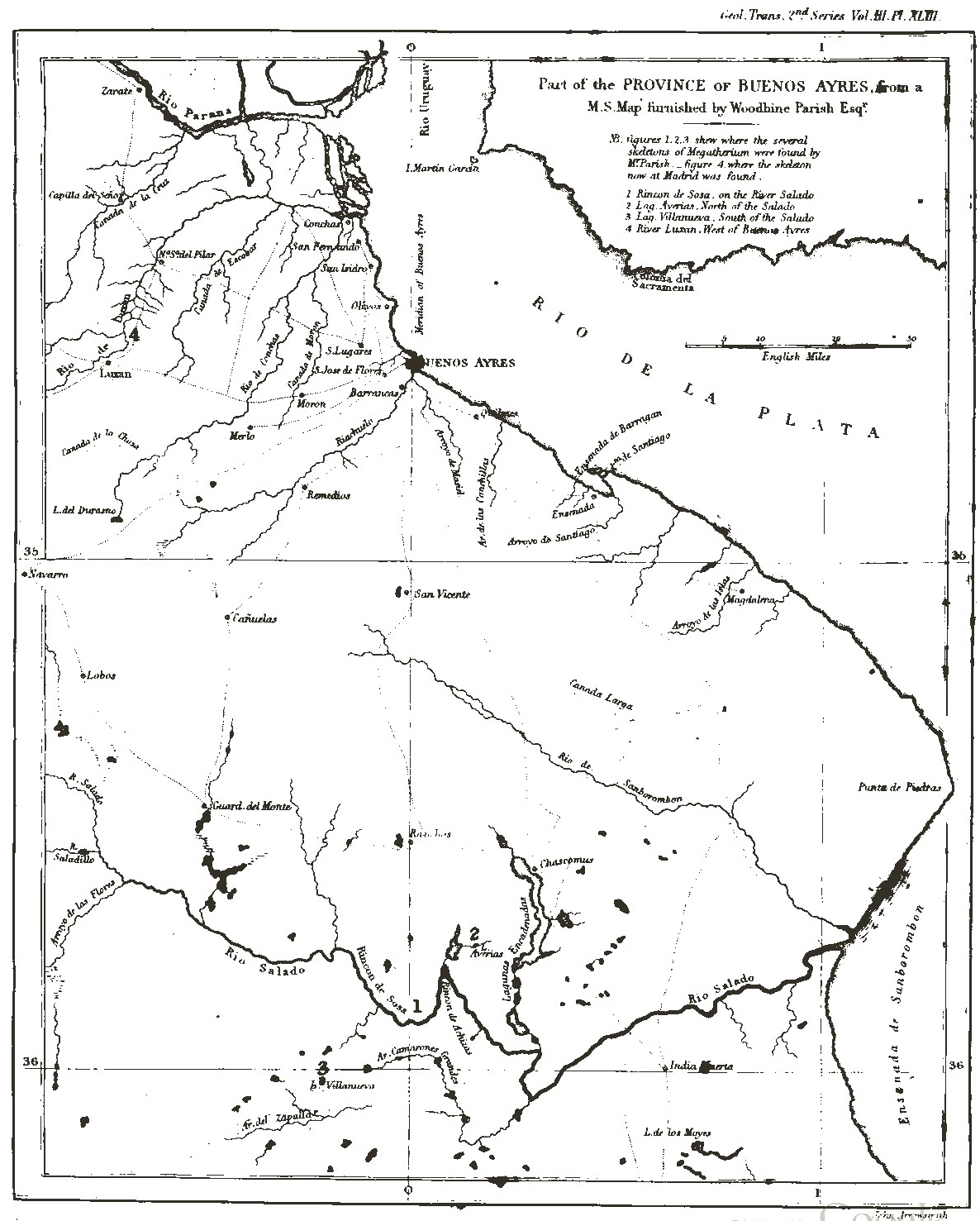
PLATE XLIII.
A copy of a portion of a Manuscript Map in the possession of Woodbine Parish, Jun., Esq., which comprehends that part of Spanish America in which those Remains of the Megatherium which have hitherto been sent to Europe were chiefly discovered.
No. 1. Denotes the situation on the River Lujan (or Luxan) whence the bones were derived which were sent to Spain in 1789 by the Marquis of Loreto, and from which were constructed the Skeleton of the Megatherium now preserved in the Royal Cabinet of Natural History at Madrid, described and figured by Don Juan Bautista Bru, and published by Don Joseph Garriga, under the title "Descripcion del Esqueleto de un Quadrúpedo muy corpulento y raro," Madrid, 1796:—and by Messrs. Pander and D'Alton under the name of "Das Riesen-Faulthier, Bradypus giganteus," Bonn, 1821.
No. 2. Rincon de Sosa, (situated in the southern part of the Map,) the property of Don Hilario Sosa, on the banks of the River Salado, the spot on which were discovered the Bones which form the subject of the present paper. Not any portion of Shell or Cuirass was found at this spot.
No. 3. The lake Las Averias, at which locality was found the most perfect example of the Cuirass, imbedded in a stratum of hard clay, at about four feet below the upper surface, together with some bones, which were exposed to view by the occasional beating of the waters against the sides of the Lake in stormy weather. The shell, when first discovered, (according to the assurances of the Peons, or country people, who accompanied the person sent by Mr. Parish to the spot,) was at least twelve feet in length, and from four to six feet in the widest or deepest part. The Bones on being taken out of the earth almost immediately mouldered away. A fragment of the pelvis was all that reached Buenos Ayres. The Skeleton was said to have appeared to be as large as that found at Señor Sosa's.
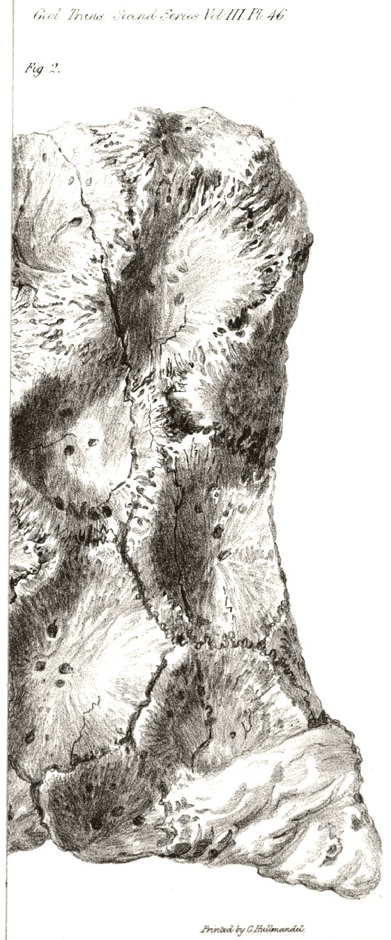
An external and internal view of a small but characteristic fragment of this Shell or Cuirass is given in Plate LXVI.
No. 4. Villanuevs. The bones found at this spot, dug out of the bed of a small rivulet, were of small size, and in a very fragile state, and crumbled to pieces on exposure to the air. Part of a jaw with one very small though nearly perfect tooth in the socket, part of a scapula, and some of the feet-bones were all that were capable of being preserved. The shell lay about a foot below the principal mass of the bones, the concave side uppermost, and resembled the section of a large cask; but would not bear to be lifted out of its bed, broke into small pieces, and crumbled to dust almost immediately.
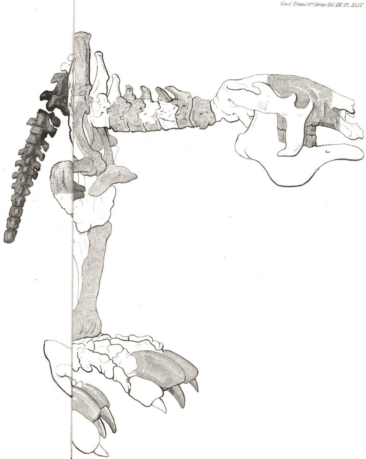
PLATE XLIV.
An outline traced from Plate I. of Messrs. Pander and D'Alton's work above-mentioned, with the intention of showing all the parts hitherto known, or supposed to be known, of this extraordinary animal, the Megatherium.
The simple outline represents the state of the skeleton, as now articulated, in the Royal Cabinet of Natural History at Madrid. Whether properly or improperly mounted, i. e. whether all the parts are of one or more individuals, whether they belong to the situation or position in which they are placed, whether all the parts
[page break]
are genuine or partly modelled, or whether parts are eked out by bones that do not belong to the part or situation in which they are now located, does not interfere with the object of the outline in this Diagram; no blame being intended to be attributed to the Articulator, who, probably, had little or no guide in such a difficult task. Upon this outline are engraved up, but in a faint degree, 1st, those parts which have been collected and preserved by Mr. Parish that also exist in the Madrid Skeleton; 2ndly, in a greater degree of strength, those parts which are preserved in the series of Bones described in this paper which are deficient in the Skeleton at Madrid; thus endeavouring to show at one view the general tenour of the Skeleton, together with all the important points hitherto determined.
PLATE XLV.
Fig. 1. represents, of the natural size, the last phalanx of a toe belonging to one of the fore feet. This has been selected as one of the smallest bones, capable of being represented on a quarto plate, that could in any degree give a just notion of the magnitude of the creature to which it appertained; and by comparison with the same bone in situ in Plate XLIV. it cannot fail of answering that intention better than the most correct description, or minute detail of admeasurement.
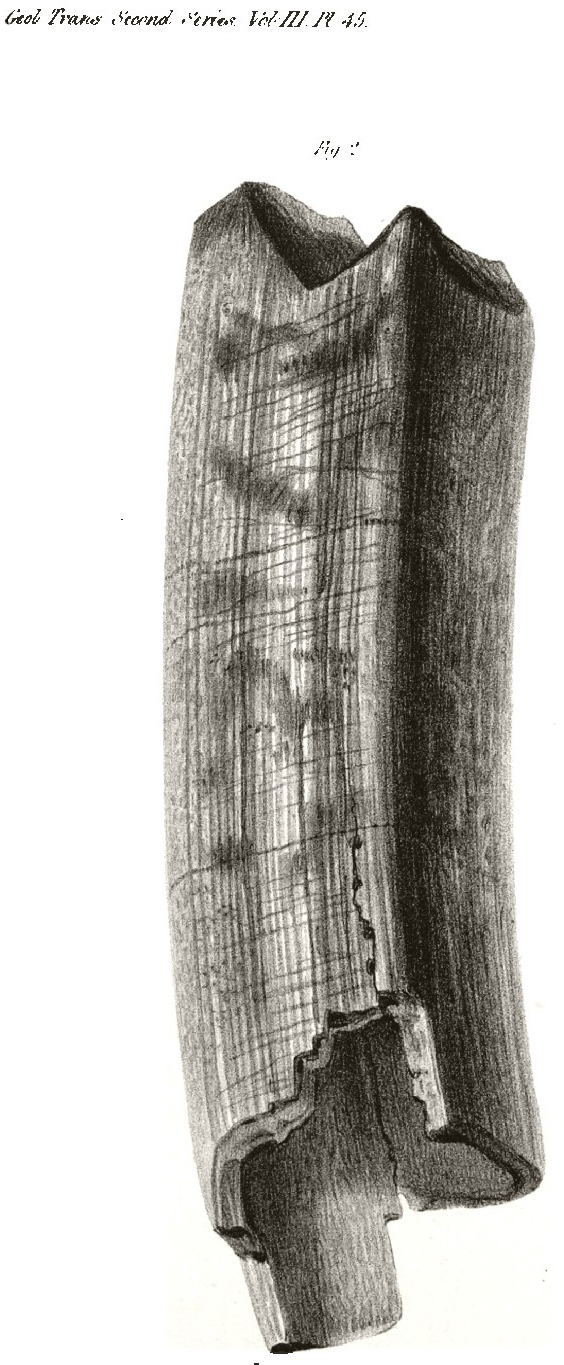
Fig. 2. represents, also of the natural dimension, a molar tooth or grinder, of which class all the teeth of the animal consist. This tooth was selected as one of the largest and most entire among those discovered; but although imperfect as regards its entire length (of which it is about two inches minus at its lower extremity, a fact determined by other specimens, less perfect in other respects), it shows more of its real form and structure than a perfect example could have permitted from any one point of view. This figure shows the inclination of the four facets of which the grinding or cutting surface of the tooth is composed,—the curved and slightly convex form of the anterior part of its body,—its flat side slightly depressed in the centre, and its flat and somewhat concave posterior surface. The fractured lower part permits a view of its square hollow cavity, which contained the vascular pyramidal pulp, on and from which the tooth was continually growing and projected upwards, in proportion as its grinding surface was worn away by attrition.
PLATE XLVI.
Two views of a small fragment of the shell or cuirass discovered at Las Averias, and described at No. 3. Plate XLIII., represented of the natural size. Although a considerable number of pieces of this armour have been preserved,—as many, perhaps, as would cover a space of five feet square,—it was difficult to select a portion sufficiently perfect on both surfaces to show its structure satisfactorily. A great part of this covering is incrusted on both surface by a very dense calcareous cement, the removal of which always produced more or less injury to the fragile surface. In this example, however, both surfaces are sufficiently perfect to show the relative size and number of tesseræ of which it is composed, and their forms, which are generally irregular hexagons. They are united to each other by indented sutures.
Fig. 1. The external surface.
Fig. 2. The internal surface.
[page break]
[page break]
[page break]
[page break]
[page break]
[page break]