[page i]
CATALOGUE
OF
MARINE POLYZOA
IN
THE COLLECTION
OF THE
BRITISH MUSEUM.
PART I.
CHEILOSTOMATA (PART).
LONDON:
PRINTED BY ORDER OF THE TRUSTEES.
1852.
[page ii]
PRINTED BY TAYLOR AND FRANCIS,
RED LION COURT, FLEET STREET.
[page iii]
NOTICE.
IN the present Catalogue it is intended to comprise figures and descriptions of all the species of Marine Polyzoa in the Collection of the British Museum,—and from typical specimens therein contained.
This Collection, already valuable, especially in the possession of Dr. Johnston's specimens, has recently been materially augmented by the addition of numerous Australian and other forms, many of them new, procured by Mr. J. McGillivray, on the Voyage of H.M.S. Rattlesnake. It has also been much enriched by the liberality of Charles Darwin, Esq., F.R.S., Dr. J. Hooker, F.R.S., Dr. Lyall, R. McAndrew, Esq., F.R.S., J.S. Bowerbank, Esq., F.R.S., the late W. Thompson, Esq., of Belfast, and others, who have placed their Collections at Mr. Busk's disposal, for the purposes of this Catalogue, and for the selection of the specimens required for the Museum Collection.
The Catalogue has been prepared, and the drawings made, by Mr. GEORGE BUSK, F.R.S., who has also super-
[page] iv
intended the execution of the lithography. The magnified figures have in all cases been taken from nature, and for the most part from specimens preserved in fluid, in order to ensure a more natural aspect than that afforded by dried specimens. As they have been drawn with the aid of the camera lucida, and to a scale which is given with each plate or figure, the absolute and relative proportions of the different objects will be at once obvious.
The other Parts will appear as soon as they can be prepared.
JOHN EDWARD GRAY.
British Museum,
August 1, 1852.
[page v]
LIST OF SPECIES.
Page
Class POLYZOA 1
Order I. P. INFUNDIBULATA 1
Suborder I. CHEILOSTOMATA 2
§ 1. Articulata 2
§§ 1. Uniserialaria 2
Family 1. CATENICELLIDÆ 2
Gen. 1. Catenicella 3
α. Fenestratæ. 1. C. lorica 6
2. C. ventricosa 7
3. C. hastata 7
4. C. aurita 8
5. C. amphora 8
6. C. plagiostoma 8
7. C. cribraria 9
8. C. margaritacea 9
β Vittatæ. 9. C. formosa 9
10. C. perforata 10
11. C. ringens 10
12. C. elegans 10
13. C. cornuta 11
14. C. umbonata 11
15. C. gibbosa 12
16. C. taurina 12
γ. Simplices. 17. C. carinata 12
Gen. 2. Alysidium 14
1. A. parasiticum 14
2. A. lafontii 14
[page] vi
Gen. 3. Calpidium 14
1. C. ornatum 15
§§ 2. Bi-multiserialaria 15
Fam. 2. SALICORNARIADÆ 15
Gen. 1. Salicornaria 16
1. S. farciminoides 16
2. S. gracilis 17
3. S. tenuirostris 17
4. S. malvinensis 18
Gen. 2. Nellia 18
1. N. oculata 18
2. N. simplex 19
Fam. 3. CELLULARIADÆ 19
Gen. 1. Cellularia 19
1. C. cuspidata 19
2. C. peachii 20
3. C. ornata 20
Gen. 2. Menipea 20
1. M. fucgensis 21
2. M. ternata 21
3. M. cirrata 21
4. M. triseriata 22
5. M. multiseriata 22
6. M. patagonica 22
Gen. 3. Scrupocellaria 23
1. S. cervicornis 24
2. S. diadema 24
3. S. scrupea 24
4. S. macandrei 24
5. S. cyclostoma 24
6. S. ferox 25
7. S. scruposa 25
Gen. 4. Canda 26
1. C. arachnoides 26
2. C. reptans 26
Gen. 5. Emma 27
1. E. crystallina 28
2. E. tricellata 28
[page] vii
§ 2. Inarticulata seu continua 28
§§ 1. Uniserialaria 28
Fam. 4. SCRUPARIADÆ 28
Gen. 1. Scruparia 28
1. S. chelata 29
Gen. 2. Hippothoa 29
1. H. catenularia 29
2. H. divaricata 30
3. H. patagonica 30
Gen. 3. Ætea 30
1. Æ. anguina 31
2. Æ. dilatata 31
3. Æ. ligulata 31
4. Æ. truncata 31
Gen. 4. Beania 32
1. B. mirabilis 32
2. B. australis 32
Fam. 5. FARCIMINARIADÆ 32
Gen. 1. Farciminaria 32
1. F. aculeata 33
Fam. 6. GEMELLARIADÆ 33
Gen. 1. Gemellaria 34
1. G. loricata 34
Gen. 2. Didymia 35
1. D. simplex 35
Gen. 3. Dimetopia 35
1. D. spicata 35
2. D. cornuta 35
Gen. 4. Notamia 36
1. N. bursaria 36
Fam. 7. CABEREADÆ 37
Gen. 1. Caberea 37
1. C. rudis 38
2. C. boryi 38
3. C. hookeri 39
4. C. lata 39
[page] viii
Gen. 2. Amastigia 40
1. A. nuda 40
Fam. 8. BICELLARIADÆ 41
Gen. 1. Bicellaria 41
1. B. ciliata 41
2. B. gracilis 42
3. B. grandis 42
4. B. tuba 42
Gen. 2. Halophila 43
1. H. johnstoniæ 43
Gen. 3. Bugula 43
1. B. neritina 43
2. B. flabellata 44
3. B. avicularia 45
4. B. plumosa 45
5. B. dentata 46
6. B. murrayana 46
Fam. 9. FLUSTRADÆ 46
Gen. 1. Flustra 47
1. F. foliacea 47
2. F. papyracea 48
3. F. truncata 48
4. F. octodon 49
5. F. denticulata 49
Gen. 2. Carbasea 50
1. C. papyrea 50
2. C. pisciformis 50
3. C. armata 50
4. C. cribriformis 51
5. C. dissimilis 51
6. C. episcopalis 52
7. C. bombycina 52
8. C. ovoidca 52
9. C. elegans 53
10. C. indivisa 53
Gen. 3. Diachoris 53
1. D. crotali 54
2. D. magellanica 54
3. D. inermis 54
[page break]
CATALOGUE
OF
MARINE POLYZOA.
Class POLYZOA.
Polyzoa, J. V. Thompson, Zool. Research. Mem. v. 92 (1830); J. E. Gray, Syn. Brit. Mus. 133; Johnst. Hist. Brit. Zooph. ed. 2. 253.
Bryozoa, Ehrenberg, Symbolœ Physicœ (1831); Corall. des Roth. Meer. 153; Jones, Anim. Kingd. 107, 117; Owen, Lect. 93, 101.
Molluscan Zoophytes s. Zoophyta Ascidioida, Johnst. Mag. Zool. & Bot. i. 448; Couch, Corn. Faun. iii. 84.
Les Bryozoaires, Audouin and Milne-Edwards, Lam. Anim. s. Vert. ed. 2. ii. 104.
Ciliobrachiata, Farre, Phil. Trans. 1837.
Polypes tuniciens, M. Edwards, Mem. 16.
Polypiaria (pars), Blainv. Dict. Sci. Nat. 1830, lx. 364.
Order I. POLYZOA INFUNDIBULATA.
Polypiaria infundibulata, P. Gervais, Ann. d. Sc. Nat. (1837) vii. 79.
B
[page] 10
elliptical, sublateral. Surface in front covered with minute acuminate papillæ
Catenicella formosa, Busk, Voy. of Rattlesn. i. 360.
Hab. Swan Island, Banks' Strait.
Colour light plumbeous. Parasitic upon C. margaritacea. The cells are the largest of any in the vittate division, and very regular and uniform in size and outline. The more distinctive characters are taken from the comparatively broad vittæ, and the flat or cupped upper surface of the avicularia, which are usually continued downwards into a prominent ridge or ala.
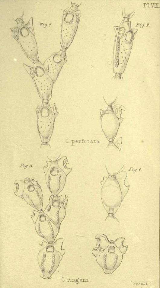
10. CATENICELLA PERFORATA. Pl. VIII. figs. 1, 2.
Cells elongated oval. Avicularian processes large, perforated at the base or by several openings. Vittæ long, wider below, lateral. Surface in front papillose.
Hab. New Zealand, Hooker, Lyall, Darwin. Tasmania, Hooker.
11. CATENICELLA RINGENS. Pl. VIII. figs. 3, 4.
Cells ovoid or subglobular. Avicularia usually very unequal, the larger one gaping. Vittæ, anterior, broad. Surface in front smooth.
Hab. New Zealand, Dieffenbach. Algoa Bay.
Differs from C. elegans, with some forms of which it might be confounded, in the absence of acuminate papillæ on the anterior surface, and in the comparatively greater size and peculiar gaping aspect of the avicularia, or not unfrequently of one of them.
12. CATENICELLA ELEGANS. Pl. IX.
Cells clongated, ovoid. Avicularia large and projecting, without any superior appendage. Vittæ narrow, sublateral. Surface in front papillose.
? Eucratea Contei, Audouin, Expl. i. 242; Savig. Eg. pl. 13. f. 1.
Catenicella Savignyi?, Blainv. Man. d'Act. 462. pl. 78. f. 1.
Catenicella elegans, Busk, Voy. of Rattlesn. i. 361. t. l. f. 2.
Hab. Bass' Strait, 47 fathoms; Port Cooper, Banks' Peninsula; Algoa Bay; Port Dalrymple.
A delicate and beautiful parasitic species; the branches slender and spreading; colour white and very transparent; cells regular, and uniform in size and shape. There appears to be little or no difference between the Australian and South African species; in the latter, however, the vittæ are usually much longer, extending upwards as high as the mouth.
[page] 18
Parasitic. Branches shorter and thicker than in the preceding species. In the shape of the area they are much alike, but in S. tenuirostris, in some cells, and occasionally throughout the greater part of an internode, the area differs widely from the more usual form. It is much expanded and arched above. In this case there is usually a considerable-sized perforation above the mouth of the cell, as occurs not unfrequently also in S. farciminoides. These indicate the situation of the immersed ovicells. The avicularium affords an excellent character between these otherwise not readily distinguishable forms.
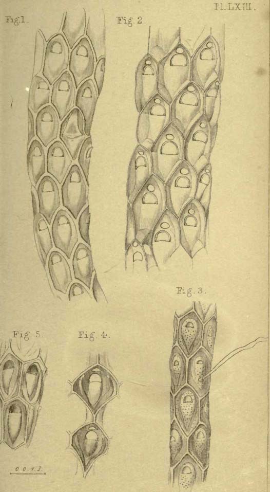
4. SALICORNARIA MALVINENSIS. Pl. LXIII. figs. 1, 2; Pl. LXV. (bis) fig. 1.
Front of cell arched above, very acute below. Cells distant in the same series. Surface smooth. Avicularium replacing a cell, rostrum immersed, mandible wide, large, triangular, pointed.
Hab. Falkland Islands, S. Patagonia, Darwin.
Readily Distinguishable by the perfect smoothness of the surface and uniformly arched form of the area above, as well as by the form of the avicularium.
2. NELLIA.
Front of cell convex, with a distinct raised border; a large aperture. No avicularia. Ovicells——?
Salicornaria, part., Busk, Voy. of Rattlesn. i. 367. Nellia, Busk, MSS.
Although evidently closely allied to Salicornaria, this genus would seem to be sufficiently distinct in the form of the front of the cell and in the complete absence of avicularia. The ovicells have not been noticed, and are probably deeply immersed. In the small number of series of the cells and their conformation it approaches the next genus, Cellularia.
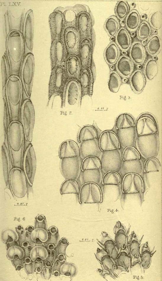
1. NELLIA OCULATA. Pl. LXIV. fig. 6; Pl. LXV (bis) fig. 4.
Outline of cell rounded above, not contracted downward, truncate below. Two raised hollow, perforated, papilliform processes below the aperture. Aperture elliptical. Cells quadriserial.
Salicornaria dichotoma, Busk, op. cit. i. 367.
Hab. Prince of Wales Channel, Torres' Strait, 9 fathoms.
Forms small crowded tufts from one to two or three inches high; branches very uniform in length, and thence very regularly forked or dichotomous.
[page] 21
In the aberrant species above referred to the cells are numerous in each internode, usually tri-or multiserial. The genus appears to enjoy a wide geographical range, occurring from the arctic circle in a species not here described, to the southern points of South America and of Africa.
a. Operculatæ. Cells with a pedunculate operculum protecting the aperture. Tricellaria.
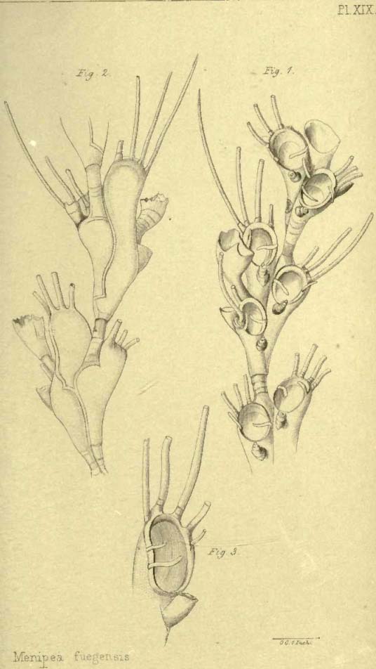
1. MENIPEA FUEGENSIS. Pl. XIX.
Cells elongated, three in each internode. Operculum simple, acicular, curved; three to four spines on the upper border. Anterior avicularium single.
Hab. Tierra del Fuego, low water, Darwin; Falkland Islands, Hooker.
2. MENIPEA TERNATA. Pl. XX. figs. 3, 4, 5.
Cells elongated, much attenuated downwards, three in each internode. Operculum expanded, entire; two spines on the upper margin. Anterior avicularium single.
Cellaria ternata, Ellis and Soland. Zooph. 30.
Sertularia ternata, Turt. Gmel. iv. 687.
Crisia ternata, Lamx. Corall. Flex. 61.
Tricellaria ternata, Flem. Brit. Anim. 540; Blainv. Act. 458; Gray, Brit. Rad. 113.
Cellularia ternata, Johnst. Hist. Brit. Zooph. ed. 2. p. 335. t. 59.
Hab. Britain.
β. Inoperculatæ. Without a pedunculate operculum. Menipea.
3. MENIPEA CIRRATA. Pl. XX. figs. 1, 2.
Cells pyriform, constricted below, six in each internode, one of the lower usually more or less aborted; usually one large lateral avicularium to each internode; three marginal spines very long and strong; anterior avicularium single, its upper border toothed.
Cellaria cirrata, Ellis and Soland. Zooph. 29. t. 4. fig. d, D.
Cellaria crispa, Pall. Elench. Zooph. 71.
Sertularia crispa, Gmel. Syst. Nat. ed. 13. 3860, and Sertularia cirrata, ib. 3862.
Tubularia cirrata, Esper, Pf. T. t. 7. fig. 1-3; Seba, Thesaur. iii t. 101. no. 8.
Menipea cirrata, Lamx. Exposit. p. 7. pl. 4. fig. D, D1; Krauss, Cor. und Zooph. d. Süds. p. 32.
Hab. South Africa.
[page] 22
4. MENIPEA TRISERIATA. Pl. XXIII. figs. 2, 3, 4.
Cells oblong, rectangular (behind), bi-triserial, numerous. Aperture oval, pointed below, and there partially filled in by a granulated expansion; one or two marginal spines on each side above. Ovicell rounded, cucullate, smooth; lip of opening entire.
Hab. South Africa.
5. MENIPEA MULTISERIATA. Pl. LX.
Cells oblong, rectangular, slightly constricted at the waist (behind); multiserial, numerous. Aperture oval, partially filled in by a granulated expansion below; a marginal spine on each side above. Ovicell large, square, with a strong and long, ascending, central umbo in front; the front lip of the opening emarginate; a radical tube inserted into the lower part of each marginal cell behind.
Hab.——? B. M. (an var. prec.)?
Although here placed under another name, there is perhaps little doubt but that this form is merely a variety of the preceding. The difference in the form of the lower part of the aperture, which in M. triseriata is described as pointed, and which in the present species usually appears square or rounded, seems to be owing to the encroachment upon it of the large ovicell. The central umbo in front of that organ, though at first sight a strongly characteristic diagnostic mark, may, as frequently happens, be owing to local conditions. It is a very common thing, especially among the Lepraliœ, that a boss-like projection of a similar kind should be thrown out on the front of the cell or of the ovicell, and usually upon both; and it is not improbable that the umbo on the ovicell in M. multiseriata may be of the same kind,—a protective spine.
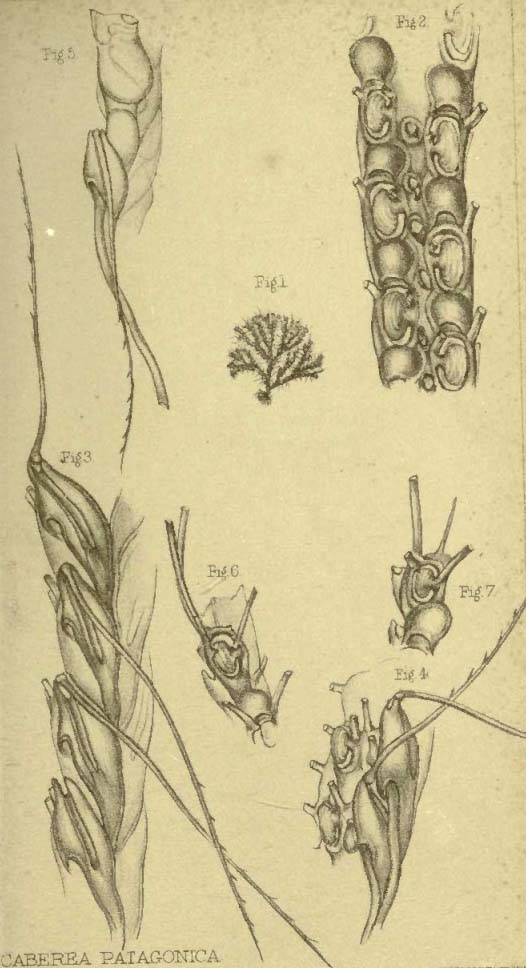
6. MENIPEA PATAGONICA. Pl. XXIII. fig. 1; Pl. XXV.; Pl. XXVI. figs. 1, 2.
Cells elongated, slightly narrowed below, six in each internode; mouth oval, simple; a very large and long spine on the upper and outer angle, below which is a sessile avicularium; a single spine on the inner edge of the aperture. Anterior avicularium single, small.
Hab. Falkland Islands, Hooker; Darwin. Port Desire, Patagonia, Darwin.
In Pl. XXV. this species is inadvertently named Cellularia. It is species very variable in the size and form of the cells, and several figures therefore of its various forms have been given. In
[page] 23
Pl. XXIII. fig. 1, is shown the mode in which the polyzoary seems to originate in a single cell, which is attached by a corneous tube to some foreign base. And in Pl. XXVI. fig. 2, is shown a curious cup-shaped appendage attached by means of a similar tube to the bottom of a cell.
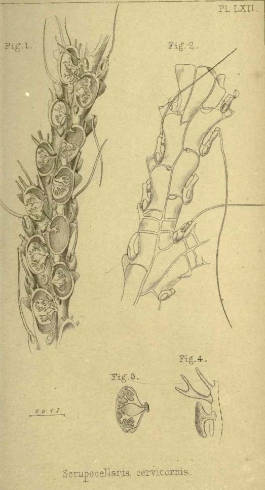
3. SCRUPOCELLARIA.
Cells rhomboidal, with a sinus on the outer and hinder aspect; each furnished with a sessile avicularium at the upper and outer angle, and with a vibraculum placed in the sinus on the outer and lower part behind. Aperture oval or subrotund, spinous above, with or without a pedunculate operculum. Cells biserial and numerous in each internode.
Scrupocellaria, Van Beneden, Recherch. 43; Gray, List of Brit. Rad. B. M. III.
Bicellaria, sp. Blainv. 1830.
Cellularia, sp. Pallas; Flem.
Cellaria, sp. Soland.; Lamk. 1816.
Scruparia, sp. Oken, Lehrb. Nat. 90, 1816.
This natural genus is characterized more particularly by the presence upon each cell of a sessile avicularium, seated, or in fact forming the upper and outer angle, and of a vibraculum placed on the back of the cell. The cells in some species are provided with a pedunculate operculum, by which it is intended to designate a process, which arising by a short tube from the anterior wall of the cell, immediately beyond the inner margin of the opening, projects forwards and bends over the front of the cell, expanding into a variously-formed limb, and serving as protection to the mouth of the cell in front. The cavity of the tube by which the process arises, becomes, in the expanded portion, continuous with variously disposed grooves or channels which terminate at the edges of the operculum. This organ affords excellent specific characters (not in this genus alone). Besides the sessile avicularia above noticed, many species of this genus also possess avicularia of another kind, and which are placed on the front of the cell below the opening and towards the inner side, or in other words, towards the middle line of the branch. In this genus, in all those species in which the second avicularium occurs, each individual cell is provided with one. This additional avicularium appears to be composed of a flexible material, and it is very easily broken off, so that in many instances, perhaps throughout an entire specimen, the organ itself may be wanting, although its position is clearly evidenced by the existence of a rounded opening in the usual situation of the organ. It is necessary to distinguish this form of flexible (if such it be) avicu-
[page] 30
Faun. iii. 101. pl. 18. fig. 5; Johnst. Hist. Brit. Zooph. ed. 2. p. 291. t. 50. figs. 9, 10; Gray, Brit. Rad. 116.
Hippothoa Elliotæ, Gray, Zool. Misc. 34.
Hab. Seas of Europe (ubique).
2. HIPPOTHOA DIVARICATA. Pl. XVIII. figs. 3, 4.
Cells remote, ovate lanceolate or fusiform; subcarinate in front; aperture small, with a notch in the lower margin. Ovicells small, superior, galeriform; branches given off at right angles, and usually in opposite pairs.
Hippothoa divaricata, Lamx. Expos. Méth. 82. t. 80. figs. 15, 16; Johnst. Hist. Brit. Zooph. ed. 2. 292. t. 51. figs. 3, 4; Audouin, Expl. i. 239; Savign. Egypt. pl. 12. f. 2 (with ovicells).
Catenicella divaricata, Blainv. D. S. N. 1830, lx. 427; Man. Act.
Hippothoa lanceolata, Gray, Zool. Misc. 35; Hassall, Ann. & Mag. Nat. Hist. vii. 366. pl. 8. figs. 5, 6; Couch, Zooph. Cornw. 43; W. Thompson, Ann. Nat. Hist. v. 252; Couch, Corn. Faun. iii. 102. pl. 18. fig. 6.
Hab. Britain. Seas of Europe.
3. HIPPOTHOA PATAGONICA. Pl. XVII. fig. 1.
Cells oval, irregularly attenuated downwards, irregularly annulate, thick; opening small, with a notch on the lower lip.
Hab. Coasts of Patagonia, Falkland Islands, Darwin.
The difference between this and the preceding one is hardly greater than what occurs in many of the Polyzoa under different circumstances of age and situation; but upon comparison of the oldest cells and the most thickened I have been able to meet with in Hipp. divaricata, I am satisfied that the distinction between that species and the present is a good one.
ÆTEA.
Cells tubular, erect, scattered, rising from a creeping fistular fibre adnate to a foreign base. Aperture terminal or subterminal.
Ætea, Lamx. Bull. Soc. Phil. 1812, char.; Gray, Brit. Rad. 133.
Anguinaria, Lamk. 1812 (no char.);Johnst. Br. Zooph. ed. 2. 292.
Falcaria β, Oken, Lehrb. Nat. 91, 1815.
The name Anguinaria appears on the plates devoted to the species of this genus, but subsequent consideration having shown the justice of recurring to Lamouroux's appellation, his term has been adopted in the text.
[page] 31
1. ÆTEA ANGUINA. Pl. XV. fig. 1.
Cells spatulate at the end; curved, ringed.
Snake Coralline, Ellis, Corall. 43. no. 11. pl. 22. fig. c, C, D.
Sertularia Anguina, Linn. Syst. ed. 10.816; Linn. Syst. 1317; Turt. Gmel. iv. 686; Berk. Syn. i. 220; Turt. Brit. Faun. 217; Stew. Elem. ii. 449; Esper, Pflanz. Sert. t. 16. figs. 1, 2; Oliv. Zool. Adriat. 291.
Cellularia Anguina, Pall. Elench. 78; Ellis, Phil. Trans. lvii. 437. pl. 19. fig. 10; Hogg's Stock. 35.
Cellaria Anguina, Ellis and Soland. Zooph. 26; Bosc, Vers, iii. 135.
Ætea Anguina, Lamx. Bull. Soc. Phil. 1812, iii. 184; Corall. 65. pl. 3. fig. 6; Exp. Méth. 9. t. 65. fig. 15; Gray, Brit. Rad. 133.
Falcaria Anguina, Oken, Lehrb. Nat. 91.
Sertularia mollis, D. Chiaje, Anim. s. Vert. Nap. iv. 147.
Anguinaria Anguina, Flem. Brit. Anim. 542; Lister, Phil. Trans. 1834, 385. pl. 12. fig. 4; Blainv. Act. 467. pl. 79. fig. 3.
Anguinaria spatulata, Lamk. Anim. s. Vert. ii. 143, ed. 2. ii. 196; Stark. Elem. ii. 439; Thompson, Ann. Nat. Hist. v. 252; Couch, Zooph. Cornw. 44; Corn. Faun. iii. 103. pl. 19. fig. 2; Johnst. Hist. Brit. Zooph. ed. 2. 290. t. 50. figs. 7, 8; Busk, Trans. Microsc. Soc. 1848, 15.
Hab. Britain, Seas of Europe, Atlantic Ocean, Antarctic Ocean, Tasmania, &c.
2. ÆTEA DILATATA. Pl. XV. figs. 2, 3.
Cells cyathiform at the apex; curved, ringed. Aperture largely dilated, suborbicular.
Anguinaria dilatata, Busk, Ann. Nat. Hist. 2nd ser. vii. 85. pl. 9. fig. 14.
Hab. Torres' Strait. Port Philip.
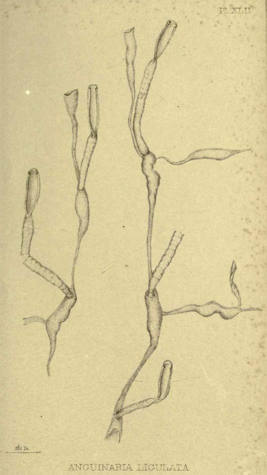
3. ÆTEA LIGULATA. Pl. XLII.
Cells very long, truncate at the extremity, straight; mouth terminal; surface not ringed; cell constricted immediately below the aperture.
Hab. Coast of Patagonia; Straits of Magellan; Darwin.
4. ÆTEA TRUNCATA.
"Cells short, straight, narrowed at their origin, extremity truncate, mouth terminal, surface punctate, not ringed."
Ætea truncata, Landsborough.
Hab. Coast of Arran; on Laminaria.
It is nearly allied to Æ. ligulata, but quite distinct.
[page] 32
4. BEANIA.
Polyzoary confervoid, subcorneous or calcareous. Cells arising one from another by a slender filiform tube given off from the lower part of the cell, which is open in front, the edges of the opening furnished with hollow spinous processes arching over the opening. Mouth terminal, with a denticle on each side.
Beania, Johnst. Brit. Zooph. ed. 2. p. 371; Gray, Brit. Rad. 96.
Though ranged among the Vesiculariadæ by Dr. Johnston, this "remarkable" genus, as he justly terms it, is clearly to be referred to the Cheilostomatous suborder. It is nearly allied on the one hand to Ætea, and on the other, through Diachoris, to the Flustradæ. As in Diachoris, the aperture occupies nearly the entire front of the erect cell. It differs however from that genus in the uniserial arrangement of the cells, and the marginal spines which defend the sides and front of the aperture.
1. BEANIA MIRABILIS. Pl. XXIV. figs. 4, 5.
Costæ seven to ten on each side.
Beania mirabilis, Johnst. Ann. Nat. Hist. v. 272; Hist. Brit. Zooph. ed. 2. p. 372. f. 69, 70.
Hab. Britain, on shells.
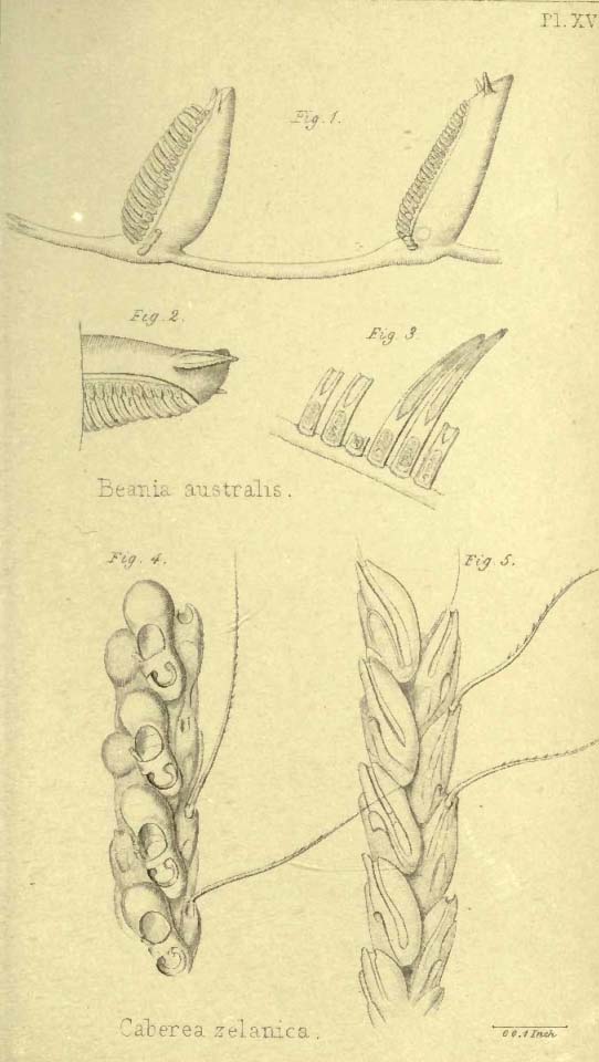
2. BEANIA AUSTRALIS. Pl. XVI. figs. 1, 2, 3.
Costæ eighteen to twenty on each side.
Hab. Coast of Patagonia; Cape Horn, Darwin, on shells and fucus.
This species sometimes appears to form a connected frond, but the cells are not so regularly interconnected as in Diachoris, and are most usually truly uniserial.
§§ 2. BI-MULTISERIALARIA. Cells disposed in a double or
multiple series.
Fam. 5. FARCIMINARIADÆ.
Cells disposed round an imaginary axis, alternate, forming cylindrical branches of an erect, dichotomously divided, continuous polyzoary.
Farciminariadæ, Busk, MSS.
1. FARCIMINARIA.
Corneous, flexible; margin of cell much raised; aperture occupying the whole front of the cell. Ovicell cucullate.
Farciminaria, Busk, MSS.
[page] 38
to allow the outlines of the cells to be seen imperfectly through them. The upper extremity of the vibraculum is bifid, and to the inner horn or tooth is articulated the "seta," and from the notch between the two horns there is continued nearly, if not quite to the inner or lower extremity of the organ, and along its upper border, a shallow groove, in which is lodged the seta when in a state of rest. In most species the seta is serrated on one side with distant teeth.
1. CABEREA RUDIS. Pl. XLVI.
Multiserial. Aperture oval, margin much thickened, with a strong projecting upturned spine on each side, in the central cells, and with three strong and long spines on the outer side, and a smaller one on the inner side in the marginal cells. Operculum spatulate, wide, entire. Each cell of the central rows with two small avicularia in front immediately below the aperture. Each marginal cell with a single large avicularium in front below the aperture. Vibracula slender, very transparent. Setæ short, not serrated.
Caberea rudis, Busk, Voy. of Rattlesn. i. 377.
Hab. Bass' Strait.
Colour dirty white. Forms a broad frondose polyzoary 2 inches or more in height. The branches all disposed in the same plane, are flat, thick, and about 1/8th inch wide, composed of 4-6 rows of comparatively small cells, which viewed behind appear lozenge or diamond-shaped, and arranged quincuncially. It is not always easy to observe with accuracy the outline of the vibracula, owing to the extreme tenuity of their walls, but the groove along the upper border is very distinct, and most usually has the seta lying in it. The avicularia on the marginal cells are very large, but not uniform in size. Along each border of the branches runs a bundle of radical tubes, the number of which diminishes as the branch ascends, owing to the circumstance that each tube terminates in the base of a vibraculum.
2. CABEREA BORYI. (Cab. zelanica, Pl. XVI. figs. 4, 5. Cab. patagonica, Pl. XXXVIII.)
Cells biserial; aperture oval, pedunculate operculum expanded principally downwards, and sometimes sending off a process to the opposite side of the aperture; a single spine on the inner side springing from the peduncle of the operculum; two marginal spines on the outer side of the aperture. Ovicell large, arcuate. Vibracula ovoid. Setæ serrated.
Crisia Boryi, Aud. Expl.; Savign. Egypt. pl. 12. f. 4.
Selbia zelanica, Gray, Dieffenb. N. Z. ii. 292.
[page] 39
Caberea zelanica, Busk, Voy. of Rattlesn. i. 378.
Hab. Cumberland Island. New Zealand, Hooker; E. Falkland Islands; S. Patagonia, 49°S.; Port St. Julian, Patagonia; Strait of Magellan, Darwin; Coast of Devon, Miss Cutler. Algoa Bay.
This appears to be one of the most generally diffused species, and it varies also considerably in some respects, according to its age and other circumstances, perhaps of depth or temperature, &c. To observe the specific characters here assigned, it is necessary to examine the younger or more perfect cells at the extremity of the branches, the older ones by continued deposition of calcareous matter being considerably altered, and also usually deprived of the spines. But the most remarkable difference is in the conformation of the pedunculate operculum. As shown in Pl. XVI. fig. 4, this process extends quite across the aperture of the cell, forming a sort of bridge, from the lower margin of which depends the expanded lamina, and this appears to be the condition in which it was figured by Savigny; whilst in Pl. XXXVIII. figs. 2, 6, 7, it will be seen that the operculum is not connected with the opposite side of the aperture, but of the more usual form. Upon sufficient examination however it will be found that both forms run insensibly into each other. The recent discovery of this species on the coast of Devonshire is of great interest. It there grows in minute tufts upon Eschara foliacea, and has probably hitherto been overlooked, owing to its resemblance to Canda reptans.
β. Inoperculatæ. No pedunculate operculum.
3. CABEREA HOOKERI. Pl. XXXVII. fig. 2.
Bi-triserial. Marginal cells with two marginal spines above and one on the inner side. Central cells with a marginal spine on each side of the aperture above. Setæ serrated.
Cellularia Hookeri, Flem. Brit. Anim. 539 (1828); Johnst. Hist. Brit. Zooph. ed. 2. 338. t. 60. f. l, 2.
Bicellaria Hookeri, Blainv. Dict. Sc. Nat. lx. 424.
Hab. Torquay, Hooker; Orkneys, E. F. Barlee.
? 4. CABEREA LATA. Pl. XLVII.
Bi-multiserial; marginal cells with a single subapical spine; central cells without marginal spines; setæ serrated.
Caberea lata, Busk, op. cit. i. 378.
Hab. Australia; New Zealand (an præced. varietas?).
[page] 40
Colour white or yellowish; forms close rounded tufts 2 to 3 inches high and wide, composed of uniform dichotomously divided branches about 1/8 th of an inch wide, and which become wider towards their truncate extremities. The vibracula are very large, and though distinctly defined, are yet sufficiently transparent to allow a view of the lozenge-shaped cells. The central rows of cells vary in number from two to five, and the cells composing them are arranged with extreme regularity. The marginal rows are placed in a plane posterior to the central, and the cells of which they are composed are widely different from the central.
It is not easy to distinguish the narrower forms of this species from Caberea hookeri, and they may not improbably really belong to one and the same species, differing only in consequence of the difference in the localities in which they are found. The warmer latitudes of New Zealand and Australia may readily be supposed to produce a more luxuriant growth, and consequently wider and stronger branches of the polyzoary. But there are other differences, which though less obvious, would better serve to indicate a specific distinction between the two forms. In Cab. hookeri there is a large tubular spine on each side of the mouth in the lateral cells, and each of the central cells, or nearly so, are furnished with an anterior avicularium, below the aperture and to one side. The lateral avicularium also of the marginal cells is much larger.
2. AMASTIGIA (α priv.,µάστιξ).
An avicularium to about each three cells on the back of the branches (no vibracula).
In this genus the vibracula on the back of the branches are replaced by avicularia; but it is to be remarked that in these avicularia, contrary to what usually obtains in those organs, the moveable mandible, when closed, points downwards; in this respect resembling the seta of the vibraculum, with which it is in fact strictly homologous.
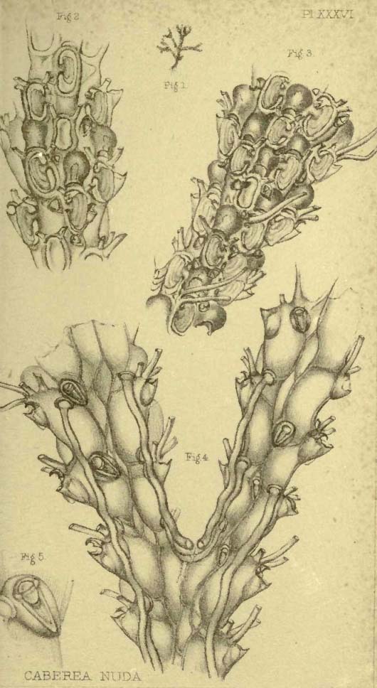
1. AMASTIGIA NUDA. Pl. XXXVI.
Cells bi-quadriserial; posterior avicularia small, the mandible pointing downwards. A lateral and anterior avicularium to each lateral cell: an anterior one to each of those in the central rows. Aperture oval, with a broad pedunculate operculum and two spines on each side above.
Caberea nuda, Busk, MSS. t. 36.
Hab Tierra del Fuego, Darwin.
[page] 52
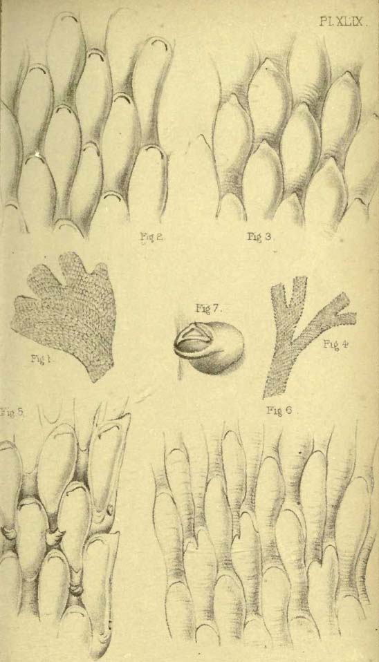
6. CARBASEA EPISCOPALIS. Pl. XLVIII.figs. 1, 2; & Pl. LV. fig. 3.
Cells pyriform, cylindrical or barrel-shaped, back marked with transverse rugæ. Aperture circular superior. Ovicells lofty, keeled. Avicularia 0.
F. pyriformis? Lamx. Pol. Flex. 103. pl. 1. fig. 4; Blainville, Man. d' Act. 451; Lamk. An. s. Vert. ii. 221; Busk, Voy. of Rattlesn. i. 379.
Hab. Bass' Strait, 45 fathoms.
Sometimes small and parasitic, upon Sertularians and Polyzoa; sometimes independent, then of large growth, forming dichotomously divided fronds, with strap-shaped, truncate, unequal segments. From its general resemblance to Lamouroux's figure, it is not improbable that this may be his F. pyriformis; but as it is impossible to determine this with certainty, either from his figure or description, (which are equally applicable to several other species,) it has been thought better to give it a new designation. The one employed is suggested by the form of the ovicells, which bear a close resemblance to a bishop's mitre.
7. CARBASEA BOMBYCINA. Pl. XLVIII. figs. 4, 5, 6, 7.
Cells pyriform, cylindrical, smooth. Aperture small, circular, superior; a lunate pore in the front of the cell a short distance below the aperture; one to four perforations, in a series on either side of the cell, above and in front. Ovicell marked with radiating lines. Avicularia 0.
? F. bombycina, Ellis and Soland. Zooph. 14. pl. 4. fig. B; Linn. Syst. Nat. ed. 13. 3828. no. 9; Bosc. Vers, 117; Lamx. Hist. Pol. Flex. 103. no. 196; Exp. Méth. 3. t. 4. fig. B; Lamk. Ann. s. V. ii. 220. 2nd ed.; Krauss, Z. d. Südsee, 35.
Hab. Algoa Bay, Mossel Bay, South Africa.
The figure of F. bombycina, given by Ellis and Solander, bears a sufficiently near general resemblance to the species here designated C. bombycina, to render it probable that they may be identical, though this is by no means certain. Considering the locality whence the present species is derived, it may not perhaps be unlikely that it represents that intended by Ellis, when he says (speaking of his F. bombycina, which came from the Bahama Islands), "I have some elegant specimens from the East Indies that approach very near to this kind."
8. CARBASEA OVOIDEA. Pl. XLIX. figs. 5, 6, 7.
Cells elongated, slightly contracted below. Aperture oval, two-
[page] 53
thirds the length of the cell, velum very convex. Ovicell—? Avicularium 0.
Hab. S. Patagonia, Darwin.
Margin of frond divided by shallow notches into small rounded lobes. The specific name is derived from the regularly oval form of the aperture, which is filled in by a very convex transparent velum.
9. CARBASEA ELEGANS. Pl. LIV. figs. 6, 7; & Pl. LVI. fig. 3.
Cells oblong. Aperture nearly as long as the cell, truncate or square below. Surface behind smooth. Ovicell—? Avicularium 0.
Hab. Tasmania.
The cells in this species most nearly resemble those of Flustra papyracea: they are however less linear and more rounded at the top, and not so long in proportion to their width. The little filling-in of the aperture at bottom affords a distinctive character, as does the absence of avicularia, were the generic difference between the two overlooked.
10. CARBASEA INDIVISA. Pl. LVIII. figs. 3, 4.
Frond semicircular, undivided, subplicated; cells oblong, surface behind granulated. Ovicells—? Avicularia 0.
Hab. New Zealand, Hooker.
3. DIACHORIS.
Cells disjunct, each connected with six others by tubular processes; frond sometimes partially adnate and decumbent.
Diachoris, Busk, Voy. of Rattlesn. 382.
The mode of arrangement and interconnexion of the cells in this genus is remarkable and highly interesting. It represents, in fact, a dissected Flustra. The cells are disposed in linear parallel series, and those of two contiguous series are alternate with respect to each other. Each cell is connected with one at either end in the same linear series by a rather wide, short tubular prolongation, and with two on each side in the contiguous series by narrower tubes; so that each cell, except in the marginal rows, is connected with six others. The species, though stated to be loosely adnate, are also capable of rising into independent erect fronds, like the other Flustradæ, and in the other form would be more correctly described as decumbent than adnate, as they are very loosely connected to the foreign base upon which they lie.
[page] 54
1. DIACHORIS CROTALI. Pl. LXVI. figs. 1, 2.
Cells erect, open in front, with straight sides; perforated on the sides and bottom; a lanceolate appendage articulated to each upper angle. Ovicell small, conical, superior.
Diachoris crotali, Busk, Voy. of Rattlesn. 382. tab. i. figs. 10, 12. Hab. Bass' Strait.
The frond, although, as explained above, not strictly adnate, as it seems to have no attachments, is usually spread loosely over other Polyzoa. There is no appearance of a moveable mandible in the lanceolate appendages, which nevertheless most probably represent avicularia. These organs are of a lanceolate form, with an elevated ridge or keel along the back, and slightly concave beneath: they project in front, slightly depending, and at the base of each is a rounded eminence.
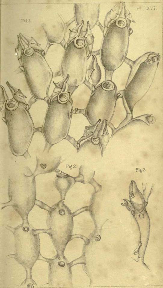
2. DIACHORIS MAGELLANICA. Pl. LXVII.
Cells semi-erect, open in front, oval; mouth circular with a thickened and raised margin. A pedunculate and articulated, capitate avicularium attached to the margin of the cell near the top on each side. Ovicell—?. (Frondose, with cells on both sides, and also loosely adnate.)
Hab. Straits of Magellan, Darwin; New Zealand, Lyall.
3. DIACHORIS INERMIS. Pl. LXXII.
Cells decumbent, boat-shaped, entirely open; two short marginal spines on each side near the top. Ovicell—? Avicularium—?
Hab. New Zealand, Lyall. Straits of Magellan, Darwin.
This species approaches very nearly to a Membranipora, and from the total absence of any moveable appendages, might perhaps be regarded as a type of a distinct genus: as, however, it is a solitary instance of the form, and agrees in the structure of the polyzoary with the above two species, it has been associated with them and not with the Membraniporidæ.
[page break]
DESCRIPTION OF PLATES.
| PLATE | ||
| I. | Fig. 1, | Catenicella lorica, p. 6, natural size; fig. 2, front; fig. 3. back. |
| II. | Fig. 1. | Catenicella ventricosa, p. 7, front; fig. 2, back. |
| Fig. 3. | Catenicella hastata, p. 7, front; fig. 4, back. | |
| III. | Fig. 1. | Catenicella ventricosa, p. 7, (var.) front; fig. 2, back; fig. 3, ovicell. |
| Fig. 4. | Catenicella ventricosa, p. 7, (var. maculata) front; fig. 5, back. | |
| IV. | Fig. 1. | Catenicella aurita, p. 8, front; fig. 2, back; fig. 3, ovicell. |
| Fig. 4. | Catenicella amphora, p. 8, front; fig. 5,back. | |
| V. | Fig 1. | Catenicella plagiostoma, p. 8, front; fig. 2, back. |
| Fig. 3. | Catenicella cribraria, front; fig. 4, back. | |
| VI. | Fig. 1. | Catenicella margaritacea, p. 9, front; fig. 2, side view; fig. 3, back. |
| Fig. 4. | Catenicella carinata, p. 12, front; fig. 5, ovicell; fig. 6, side view. | |
| VII. | Fig. 1. | Catenicella formosa, p. 9, front; fig. 2, back. |
| Fig. 3. | Catenicella gibbosa, p. 12, front; fig. 4, back. | |
| VIII. | Fig. 1. | Catenicella perforata, p. 10, front; fig. 2, back and side views. |
| Fig. 3. | Catenicella ringens, p. 10, front; fig. 4, back. |
[page] ii
| IX. | Fig. 1. | Catenicella elegans, p. 10 (var. South Africa), front; fig. 2, back; fig. 3 (var. Australia), front; fig. 4, back. |
| X. | Fig. 1. | Catenicella cornuta, p. 11, front; fig. 2, back; fig. 3, side view. |
| Fig. 4. | Catenicella umbonata, p. 11, front; fig. 5, back. | |
| XI. | Fig. 1. | Catenicella taurina, p. 12, front; fig. 2, back; fig. 3, with ovicells; fig, 4, spines replaced by avicularia. |
| XII. | Fig. 1. | Calpidium ornatum, p. 15, natural size; fig. 2, front. |
| XIII. | Fig. 1. | Calpidium ornatum, p. 15, front view of bifurcation; fig. 2, back of cell. |
| XIV. | Fig. 1. | Alysidium lafontii, p. 14, front of cell; fig. 2, side view, showing the avicularium; fig. 3, back; fig. 4. back, at a bifurcation; fig. 5, natural size. |
| Fig. 6. | Alysidium parasiticum, p. 14, front, with an ovicell; fig. 7, back; fig. 8, side view; fig. 9, natural size. | |
| XV. | Fig. 1. | Ætea anguina, p. 31. |
| Fig. 2. | Ætea dilatata, p. 31. | |
| XVI. | Fig. 1. | Beania australis, p. 32; fig. 2, portion of cell to show the lateral processes; fig. 3, a more highly magnified view of the costœ. |
| Fig. 4. | Caberea boryi, p. 38, front; fig. 5, back. | |
| XVII. | Fig.1 | Hippothoa patagonica, p. 30. |
| Fig. 2 | Scruparia chelata, p. 29. | |
| XVIII. | Fig. 1, 2. | Hippothoa catenularia, p. 29. |
| Fig. 3, 4. | Hippothoa divaricata, p. 30. | |
| XIX. | Fig. 1. | Menipea fuegensis, p. 21, front view; fig. 2, back view; fig. 3, larger view of mouth of cell with the avicularium. |
| XX. | Fig. 1. | Menipea cirrata, p. 21, front; fig. 2, back. |
| Fig. 3, 5. | Menipea ternata, p. 21, front; fig. 4. back. |
[page] iii
| XXI. | Fig. 1. | Scrupocellaria scrupea, p. 24, front; fig. 2, back. |
| Fig. 3. | Canda reptans, p. 26, front; fig. 4, back. | |
| XXII. | Fig. 1. | Scrupocellaria ferox, p. 25, front; fig. 2, back; fig. 5, ovicell. |
| Fig. 3. | Scrupocellaria scruposa, p. 25, back; fig. 4,front. | |
| XXIII. | Fig. 1. | Menipea patagonica, p. 22, mode of origin. |
| Fig. 2. | Menipea triseriata, p. 22, front; fig. 3, back; fig. 4,ovicells. | |
| XXIV. | Fig. 1. | Scrupocellaria macandrei, p. 24, front; fig. 2, back; fig. 3,radical tube. |
| Fig. 4. | Beania mirabilis, p. 32, side view of cell; fig. 5, enlarged view of costœ. | |
| XXV. | Fig. 1. | Menipea patagonica, p. 22, front; fig. 2, 3, back. |
| XXVI. | Fig. 1, 2. | Menipea patagonica, p. 22, variety. |
| Fig. 3. | Cellularia ornata, p. 20, front; fig. 4, back. | |
| XXVII. | Fig. 1. | Cellularia cuspidata, p. 19, front.; fig. 2, back. |
| Fig. 3. | Cellularia peachii, p. 20, front; fig. 4, back; fig. 5, ovicell. | |
| XXVIII. | Fig. 1. | Scrupocellaria diadema, p. 24, front; fig. 2, back; fig. 3, ovicell. |
| Fig. 4. | Scrupocellaria cyclostoma, p. 24, front; fig. 5, back. | |
| XXIX. | Fig. 1. | Dimetopia spicata, p. 35. |
| Fig. 2. | Dimetopia cornuta, p. 35; fig. 3, ovicell. | |
| XXX. | Fig. 1. | Halophila johnstoniæ, p. 43; natural size; fig. 2, back; fig. 3, front. |
| XXXI. | Fig. 1. | Bicellaria tuba, p. 42, front; fig. 2, back, with avicularium; fig. 3, occasional mode of connexion of cells; fig. 4, ovicell. |
| XXXII. | Fig. 1. | Bicellaria gracilis, p. 42, natural size; figs. 2, 4, front; figs. 3, 5, back. |
| XXXIII. | Fig. 1. | Canda arachnoides, p. 26, natural size; figs. 2, 4, front; fig. 3, back; fig. 5, avicularia at the lower part of a branch |
[page] iv
| XXXIV. | Fig. 1. | Bicellaria ciliata, p. 41, natural size; fig. 2, front; fig. 3, back; fig. 4, ovicells; fig. 5, avicularia. |
| XXXV. | Fig. 1. | Bugula dentata, p. 46, natural size; fig. 2, front; fig. 3, back; fig. 4, side; fig. 5, avicularia. |
| XXXVI. | Fig. 1. | Amastigia nuda, p. 40, natural size; figs. 2, 3, front; fig. 4, back; fig. 5, dorsal avicularium. |
| XXXVII. | Fig. 1. | Caberea hookeri, p. 39, natural size; fig. 2, front; fig. 3, back. |
| XXXVIII. | Fig. 1. | Caberea boryi, p. 38, natural size; fig. 2, front; fig. 3, back; figs. 4, 5, side; figs. 6, 7, younger cells. |
| XXXIX. | Fig. 1. | Didymia simplex, p. 35, natural size; fig. 2, front; fig. 3, front with ovicell. |
| XL. | Fig. 1. | Emma crystallina, p. 28, natural size; fig. 2, front; fig. 3, back. |
| XLI. | Fig. 1. | Emma tricellata, p. 28, front; fig. 2, back. |
| XLII. | Ætea ligulata, p. 31. | |
| XLIII. | Fig. 1. | Bugula neritina, p. 43, natural size; fig. 2, front; figs. 3, 4, back; figs. 5, 6, ovicells. |
| XLIV. | Fig. 1. | Bicellaria grandis, p. 42, natural size; fig. 2, front; fig. 3, back. |
| XLV. | Fig. 1. | Notamia bursaria, p. 36, natural size; fig. 2, front; fig. 3, back; fig. 4, Diagram to represent the mode of connexion of the cells. |
| Fig. 5. | Gemellaria loricata, p. 34, natural size; fig. 6, back and side. | |
| XLVI. | Fig. 1. | Caberea rudis, p. 38, natural size; fig. 2, front; fig. 3, back. |
| XLVII. | Fig. 1. | Caberea lata, p. 39, natural size; fig. 2, front; fig. 3, back. |
| XLVIII. | Fig. 1. | Carbasea episcopalis, p. 52, natural size; fig. 2, front; fig. 3, back. |
[page] v
| XLVIII. | Fig. 4. | Carbasea bombycina, p. 52, natural size; fig. 5, front; fig. 6, back; fig. 7, ovicells. |
| XLIX. | Fig. 1. | Carbasea armata, p. 50, natural size; fig. 2, front. |
| Fig. 3. | Flustra denticulata (var. inermis), p. 49, natural size; fig. 4, magnified. | |
| Fig. 5. | Carbasea ovoidea, p. 52, natural size; fig. 6, front; fig. 7, back. | |
| L. | Fig. 1. | Carbasea papyrea, p. 52, natural size; fig. 2, front; fig. 3, back. |
| Fig. 4. | Carbasea dissimilis, p. 51, natural size; fig. 5, front; fig. 6, back; fig. 7, avicularium. | |
| LI. | Fig. 1. | Bugula flabellata, p. 44, natural size; fig. 2, front; fig. 3, avicularium (more magnified). |
| LII. | Bugula flabellata, p. 44, back. | |
| LIII. | Fig. 1. | Bugula avicularia, p. 45, natural size; fig. 2, front; fig. 3, back; fig. 4, avicularium. |
| LIV. | Fig. 1. | Bugula plumosa, p. 45, natural size; fig. 2, front; fig. 3, back; fig. 4, ovicell; fig. 5, avicularium. |
| Fig. 6. | Carbasea elegans, p. 53, front; fig. 7, back. | |
| LV. | Fig. 1. | Carbasea pisciformis, p. 50, front; fig. 2, back. |
| Fig. 3. | Carbasea episcopalis, p. 52, front (without ovicells). | |
| Figs. 4, 5. | Flustra foliacea, p. 47. | |
| Figs. 6, 7. | Flustra papyracea, p. 48. | |
| LVI. | Figs. 1, 2. | Flustra truncata, p. 48. |
| Fig. 3. | Carbasea elegans, p. 53. | |
| Fig. 4. | Flustra octodon, p. 49. | |
| Fig. 5. | Flustra foliacea, p. 47. | |
| Fig. 6. | Carbasea pisciformis, p. 50. | |
| Fig. 7. | Flustra denticulata, p. 49. | |
| LVII. | Fig. 1. | Flustra denticulata, p. 49, portion of frond; fig. 2, a single cell with avicularium more highly magnified. |
| LVIII. | Figs. 1, 2. | Flustra truncata, p. 48. |
| Fig. 3. | Carbasea indivisa, p. 53, front; fig. 4, back. | |
| Fig. 5. | Flustra octodon, p. 48. | |
| Fig. 6. | Membranipora telacea. Vid. Part II. |
[page] vi
| LIX. | Fig. 1. | Bugula murrayana, p. 46, front; fig. 2, back. | |
| LX. | Fig. 1. | Menipea multiseriata, p. 22, front; fig. 2, back. | |
| LXI. | Fig. 1. | Membranipora lineata,. Vid. Part II. | |
| Fig. 2. | Membranipora membranacea. Vid. Part II. | ||
| LXII. | Fig. 1. | Scrupocellaria cervicornis, p. 24, front; fig. 2, back; fig. 3, pedunculate operculum; fig. 4, marginal spines. | |
| LXIII. | Figs. 1, 2. | Salicornaria malvinensis, p. 18. | |
| Fig. 3. | Salicornaria gracilis, p. 17. | ||
| Fig. 4. | Salicornaria tenuirostris, p. 17. | ||
| Fig. 5. | Salicornaria teuuirostris, var. a, p. 17. | ||
| LXIV. | Fig. 1-3. | Salicornaria farciminoides, p. 16. | |
| Fig. 2. | Var. (sinuosa). | ||
| Figs. 4, 5. | Farciminaria aculeata, p. 33. | ||
| Fig. 6. | Nellia oculata, p. 18. | ||
| LXV. | Fig. 1. | Nellia simplex, p. 19. | |
| Fig. 2. | Vincularia ornata. Vid. Part II. | ||
| Fig. 3. | Membranipora cyclops. | ||
| Fig. 4. | Membranipora grandis. | ||
| Fig. 5. | Membranipora galeata. | ||
| Fig. 6. | Membranipora rozieri. | ||
| LXV.(*). | Fig. 1. | Salicornaria malvinensis, p. 18, natural size. | |
| Fig. 2. | Salicornaria gracilis, p. 17, natural size. | ||
| Fig. 3. | Nellia simplex, p. 19, natural size. | ||
| Fig. 4. | Nellia oculata, p. 18, natural size. | ||
| Fig. 5. | Salicornaria farciminoides, p. 16, natural size. | ||
| Fig. 6. | Farciminaria aculeata, p. 33, natural size. | ||
| Fig. 7. | Farciminaria flexilis, natural size. Vid. Part II. | ||
| LXVI. | Fig. 1. | Diachoris crotali, p. 54, front; fig. 2, back; fig. 3, side (reduced). | |
| LXVII. | Fig. 1. | Diachoris magellanica, p. 54, front; fig. 2, back; fig. 3, side. | |
| LXVIII. | Fig. 1. | Carbasea cribriformis, p. 51. | |
| Fig. 2. | Membranipora telacea. Vid. Part II. |
[page vii]
N. B. The present Part contains about one-half of the Cheilostomatous suborder, the remainder of which will be included in a second. Owing to the rapid accumulation of materials during the progress of the Work, it has not been possible to adhere to any regular sequence in the Plates, and it has been found necessary to reserve one Plate (LXXII.), containing the figures of Diachoris inermis, for the ensuing part of the Catalogue; for the same reason also the figures of several species of Membranipora and of a species of Vincularia, to be afterwards described, appear in Plates LVIII., LXI., LXV. and LXVIII. G. B.
[page break]
[page break]
[page break]
Pl. XVII
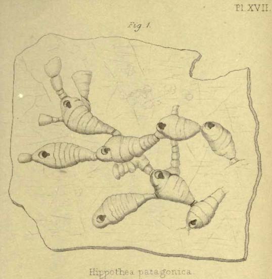
Hippothea patagonica
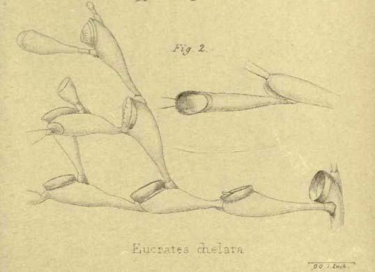
Eucratea chelata
[page break]
[page break]
[page break]
[page break]
[page break]
[page break]
[page break]
[page break]
[page break]
[page break]
[page break]