[page] 79
VI. An account of the Two Methods of Reproduction in Daphnia, and of the Structure of the Ephippium. By JOHN LUBBOCK, Esq., F. G. S. Communicated by CHARLES DARWIN, F.R.S.
Received December 22, 1856.—Read January 29, 1857.
THE genus Daphnia is a small group of freshwater Entomostracous Crustacea, which has long been celebrated for possessing the power of reproduction without the intervention of the male, and for laying two different sorts of eggs. In both cases the eggs pass from the ovary into a space or receptacle between the carapace and the back of the animal. The carapace, according to MILNE-EDWARDS, is an excessive development of an anterior, probably the mandibular segment* which encloses the whole of the posterior part of the body, which lies freely in it, as it was in a bivalve shell. This bivalve shell or carapace is widely open below and behind, but is closed along the back, and as it form there a regular arch, and does not follow the sinuous margin of the back of the animal, there is left between the back and the carapace an open space of receptacle, into which the eggs are laid, and in which they remain until the young Daphnia is sufficiently developed to be able to swim about by itself.
This "receptacle" is freely open to the surrounding water, but the eggs are prevented from falling out by a tongue-like projection, developed evidently for that purpose from the back of the animal. When, however, the young are ready for exclusion, the mother has but to depress her abdomen, and they easily slip from the receptacle into the open water. Although both the common or agamic† and the ephippial eggs are thus protected by the carapace or valves until they are hatched, there is this difference, that the agamic eggs are carried about by the animal, and shortly before the next shedding of the skin, the young which have in the meantime been hatched swim away; which for the ephippial eggs, part of the carapace is specialized into a pod-like or saddle-shaped box called the ephippium, in which, when the skin is changed, they remain for some time before being hatched.
Several of the earlier naturalists, and more recently Dr. BAIRD, in his excellent History of British Entomostraca,' have detailed observations clearly showing that the ordinary eggs are produced, and are fertile, without the intervention of the male, but the mode of production of the ephippial eggs has never been correctly described; nor
* See Ann. des Sc. Nat. 1851, p. 234.
† The agamic eggs certainly do not require impregnation, and I shall give my reason presently for believing that the male influence is necessary to the hatching of the ephippial eggs; but this is not proved, and it is therefore just possible that both sorts of eggs may be agamic.
M 2
[page] 80
has it been determined whether they also are agamic, or whether the stimulus of impregnation is necessary to bring them to maturity.
The agamic reproduction of Daphnia has not, indeed, been sufficiently appreciated in the different essays which have recently appeared on this subject, and M. DE QUATREFAGES even says, in an essay on "Les Metamorphoses*," "Des cinq class composant le sousembranchement des Annelés supérieurs, ou Annelés à pieds articulés, les insectes seuls paraissent se reproduire par généagénèse;: ou n'a du moins encore rien observé de semblable chez les Myriapodes, les Arachnides, les Crustacés, ou les Cirrhipèdes."
The fact, however, rests upon such excellent evidence, that I need not here do more than refer to the observations already mentioned, which are fully confirmed by my own experience, and which may easily be repeated by any naturalist who has still some lingering doubts upon the subject.
I propose now to describe,—
First. The male organs.
Secondly. The formation of the ephippium and the ephippial eggs.
Thirdly. The formation of the common or agamic eggs.
Finally. To collect the scattered notices of parthenogenesis among the Articulata, and to endeavour to show that eggs and buds are not bodies essentially different in their nature, and that between them no distinct line of division can be drawn.
Before, however, entering on my subject, it may perhaps be as well to detail the mode in which I carried on my observations.
My stock of Daphiæ were kept in glass vessels, holding two or three pints each, and they bred there without giving any trouble. Their food consisted of Scenedesmus obliquus and other microscopic plants, which covered the sides and bottom of the glasses with a thin green film. Daphnia Schœfferi was the species I selected, because it is the largest of the genus, and because it could be at any time easily obtained from a pond in my neighbourhood. This seemed to be important, because, as the species is generally found in horseponds, I feared that my specimens would not live long in confinement. On the contrary, however, they throve well, though in the course of a few generations they diminished considerably in size, suggesting the idea whether they might not after all be merely a gigantic variety of Daphnia pulex. This diminution of size in animals kept in vivaria, as compared with those living in their native haunts, is not confined to Daphniæ, nor does it appear, at least in this instance, to be caused by any injurious effect of confinement, because the specimens seemed very healthy and multiplied freely: perhaps, therefore, it may be connected with the known fact that insular specimens have a tendency to become smaller than continental ones of the same species.
When first taken from their native ponds, my Daphniœ were generally covered with Voticellæ, parasitic plants, &c., which materially impaired their transparency; but I was very glad to find that when they had been some time in captivity, they became cleaner and more transparent, and that this was especially the case those which, for the
* Revue des deux Mondes, 1 Avril, 1855.
[page] 81
purpose of continuous watching, I kept by themselves in tumblers. For the purposes of observation, I used on of SMITH and BECK'S ordinary glass cells, about 1/20th inch in thickness. This, being glued on to a glass slide, and a piece of thin glass placed over it, formed a cell, which held the Daphnia, and prevented it from moving, without doing it any injury. To put them into this cell I used a small metal tube, and in this manner I was able to watch the same specimen at intervals of a few hours, for several days together, and to observe gradually the formation of the eggs in the ovary, without doing any injury to my subject. To identify my specimens without fear of mistake, I placed in each tumbler a small bit of tinfoil, stamped with the number of the page in my note-book.
Until now, "the male organs of generation have never been observed*," and though ZENKER† has described the testes of various species and the generative opening, he has placed and even figured the later in probably the wrong place. Speaking of Sida crystallina, he says, "Sein Ausführungsgang mündet an der dan Füssen zugekehrten Seite des Schwanzes auf. Diese Geschlechtsöffnung variert nur sehr wenig ihre Lage durch alle Daphnoiden." Either, however, the situation of the orifice in question does differ considerably in different species, or else he is certainly wrong in the place he assigns to it.
If the tails of a male and female Daphnia (Pl. VII. figs. 6 and 7) are compared, it will be at once seen that the papillæ (a) are much larger in the male than in the female. The vas deferens will almost always be seen leading to this point, and if a mature male be slightly compressed, two clouds of minute bodies will be seen to come out in a stream, one from the papilla of each side of the body, and gradually diffuse themselves in the surrounding waters.
The convince myself that these were really spermatozoa, on the 16th and 27th of June I compressed seven males, and altogether I must have examined about thirty, all with the same result. I then compressed seven females, and neither then nor at any other time could I observe anything similar in this sex. There can, threrefore, be no doubt that these are the generative orifices, and that the minute bodies are the spermatozoa.
These latter are rod-like bodies, about ·0004 of an inch in length, and ·000125 of an inch in breadth. They are almost stationary, but if closely watched, a slight wriggling movement can be observed, and several may generally be found curved into an arched form, which, however, may perhaps not be natural, but may be caused by the action of the water. Among these rod-like spermatozoa were several round, probably spermatic, cells, in most of which two bright points could be discerned, which I presume to be the two ends of the spermatozoon.
I have in vain endeavoured to observe the penetration of the spermatozoon into the ovary. Although it is not difficult at certain seasons to find couples attached together, and to place them under the microscope, yet I have always found the male to be merely hanging on to the female by the organs provided for that purpose, and not to be at the
* BAIRD, loc. cit. p.77.
† "Physiologische Bemerkeungen über die Daphnoiden," MÜLLER'S Archiv, 1851.
[page] 82
moment engaged in a true act of coition. Moreover, the forms of the papilla, which can scarcely be called a penis, justifies the inference that the act of intromission, if any, would be short and easily interrupted. At any rate, I must leave the question as to the mode of introduction of the spermatozoa, undecided for the present. The facts to be hereafter mentioned as to the development of the ephippial egg render it probable that the impregnation takes place at an early period in the development of the ovarian egg, which, unless the act is effected, gradually disappears.
Daphniœ may, however, be found in every stage of ovarian development united to one or two males. I have found them so when the ovaries have been occupied only by nucleated ovarian masses, when the have been almost ready to escape into the receptacle, and when the ephippium has been almost mature. In these cases the coition is probably without any effect; and, indeed, though I have very seldom seen specimens with ephippia where I could find no males, and though the abundance of the males appears to vary with the number of the ephippia, yet I have often confined males and females together in the hope of being thus enables to trace out the manner of the impregnation of the ephippial egg, but without succeeding in any one instance.
Thus, on the 30th of September, two specimens which were confined together were in coition: this lasted for three-quarters of an hour after I first observed them. The female had then, at the centre of the right ovary, an agamic egg commencing, formed of a somewhat enlarged germinal vesicle, surrounded by some oil-globules and brown granules. On the evening of October 2nd, I again found these two specimens in coition, for a least three-quarters of an hour. At seven o'clock on the morning of the 3rd, I found that eggs had been deposited in the receptacle during the night, and the pair were again in coition. In the ovary were only the usual ovarian masses. On the 4th, 5th and 6th they were again in coition, and on the morning of the 7th a fresh set of eggs had been deposited in the receptacle. Again, on the 19th of September, I placed eleven females in a glass with about as many males, and watched them till the 19th of October, when two females and three males were still alive, and during that time not one ephippium was produced.
These experiments, however, and many others with similar results prove only that the mere stimulus of the presence of males is not sufficient by itself to cause the development of ephippia, but that other concurrent circumstances are necessary.
It has been stated by most writers that the males and ephippia appear only at certain season: ZENKER, however*, has stated that the males may be found all though the year, and I have myself observed both males and ephippia from the beginning of May till the end of December, with only intervals here and there of a few days. Moreover, the ephippial eggs laid in the spring, summer, and autumn, did not, as is often stated, remain undeveloped till the following spring, but at least in many cases were hatched without any great interval.
I have only once, and then not very satisfactorily, been able to see the actual deposi-
* Loc. cit.
[page] 83
tion of the agamic eggs in the receptacle, but I have several times observed the state of the ovary within a few minutes after this took place. The ovary was then as distinct as usual: it consisted of a slightly undulating tube, varying in the middle from ·0033 to ·005 in width, but contracted at both ends, where also the walls became fainter, so that neither the anterior nor the posterior termination could be observed. The anterior contracted part appeared to be empty, or at least a few irregular curved lines only were visible. Nearly opposite to the hind end of the heart was generally an elliptic ovarian mass, with a constriction in the centre of the upper margin, and containing from two to four, or at most five cells, each consisting of a large, perfectly circular nucleus, surrounded by the cells, which formed a sort of narrow border to it. The nucleus can sometimes scarcely be distinguished, but is usually slightly darker than the ovarian mass itself, and this latter than the cells. This ovarian mass occupies the whole width of the ovary, and is followed in a linear series by three or four more, all very similar to the first, all with the lower margin regularly arched, the upper constricted at or near to the centre. The longer axes of these ovarian masses, which is in the direction of the longer axis of the body, is about ·0058 in length, the shorter ·0033: the diameter of the cells varies from ·001 to ·0004. After the first three or four, the masses become smaller, two or more are abreast, and the walls gradually more indistinct: the cells also are usually smaller behind, but not regularly so, often a mass with large cell behind one with small ones. In many cases, however, the order of arrangement is not so regular, nor the masses so distinct; but they appear, as it were, heaped together, and even where the ovary is at first as described above, it gradually changes to this latter appearance. On all sides of the ovary are often scattered circular cells, containing each one or more oil-globules, but they are most numerous at the downward bend of the abdomen. A few hours, or sometimes immediately, after the deposition of the agamic eggs in the receptacle between the back and the shell, it usually happens that at the posterior part of the ovary one of the above-mentioned cells swells a little, and becoming surrounded by brown granules, may be called a germinal vesicle (Plate VI, fig. 2 g′). Other cells, sometimes two or three in number, but usually only one, are at first visible in the ovarian mass, but gradually disappear. The contents of this mass continue to grow darker, until it comes to resemble Plate VI. fig. 3 g′; after this they generally either fade away, or break up into more or less compact little balls (Plate VI. fig. 8) which gradually disappear.
The ephippial egg in these early stages differs from the corresponding state of agamic eggs in the determinate position and number, being never more than one on each side of the body; and in the constant absence of the large oil-globules, which are constantly present, and show themselves very early in the latter form of eggs.
This embryonic ephippial egg may generally be met with in those specimens which have recently moulted and laid their eggs, and indeed I should say that the cases in which it is not produced are quite exceptional, probably not more than one in five or six.
I have traced the progress of it in thirty-three instances, and in thirty it gradually disappeared again as described above; in the other three the dark granules increased
[page] 84
greatly in number, and formed finally a large, firm-looking, dark homogeneous mass, the ephippial egg. In its later stages it may easily be distinguished from the matter of the agamic eggs, by the darker colour, more regular outline, and absence of large oil-globules.
There are never more than two ephippial eggs in Daphnia, one being formed in each ovary. Prof. HUXLEY appears to have not very clearly apprehended the relation of these two ova to one another, and to the ephippium, for he compares them to two masses formed in the ephippial egg of Lacinularia*, whereas they are two distinct eggs, and the ephippium of Daphnia is not an egg-shell like the external shell of the winter eggs of Rotifers, but a portion of the carapace, and therefore analogous to the skin of the female Coccus, which acts as a protection to the eggs beneath it. During the last hours before its deposition in the ephippium, the ephippial egg appears to occupy the whole of the ovary, or rather the latter cannot be distinguished; and if at this time the ephippial egg is removed from the body and compressed, there will be seen an immense number of minute cells, some ·00083, but by far the greater number about ·0001 in diameter, and also some of the usual ovarian cells containing the nuclei, which I call germinal vesicles. These latter, however, do not, I believe, belong to the egg itself: the chorion is at this period so delicate, that I have never been able completely to isolate the egg from the other contents of the ovary; but I examined it several times as soon as it was laid, and found it to contain only the above-mentioned minute cells. The mother-cells, or those containing the germinal vesicles, could then be clearly seen in the ovary, and I believe, therefore, that the above-mentioned disappearance of the ovary is only apparent.
The most accurate account given of the ephippium is that by STRAUSS, who syas†:— "à certaine époque de l'année, notamment vers les mois de Juillet et d'Août, les valves de la femelle prennent, après la nuce, de l'opacité dans leur partie supérieure, chacune dans une étendue à peu près rectangulaire, s'étendant depuis les environs du premier segment jusqu'au sixième, et descendant jusqu'au dessous de la région des ovaires. Cette partie opaque est d'abord de couleur un peu blanchâtre: mais devenant bientôt plus fonçée, elle finit par être d'un gris noirâtre assez obscur. Sur chacune on aperçoit deux ampoules ovulaires, transparentes, placeés l'une au devant de l'autre, et formant avec celles du côté opposé deux petites capsules ovales, s'ouvrant comme une coquile bivalve. MÜLLER a nommé ces pièces opaques un ephippium, sans dire toutefois ce qu'il en pensait: et comme elles ont en effet, vu leur situation sur le dos de l'animal, quelque ressemblance avec une selle, j'ai cru devoir conserver cette dénomination, pour ne pas introduire de nouveaux termes inutiles. Cet ephippium se partage comme les valves, dont il fait partie, en deux moitiés latérales, réunies par suture le long de leur bord supérieur. Dans son intérieur on en trouve un autre semblable, mais plus petit, à bords libres, et dont les deux moitiés jouent en charnière une sur l'autre."
This account is very accurate: STRAUSS expressly states that the ephippium is a part of the carapace, a fact which has been miscomprehended by subsequent writers. Thus, Dr. BAIRD says (p. 85) that a green matter passes from the ovaries into the receptacle,
* Loc, cit. p. 14.
† Ann. de Musée, 1819, vol. v. p.415.
[page] 85
"and there spreading, forms the ephippium." Professor HUXLEY compares the ephippium with the outer coat of the winter egg of Lacinularia, which is a product of the ovary. Prof. MILNE-EDWARDS says*, "On voit alors (vers la fin l'été) se former dans la cavité ovifère, à la face interne de chaque valve, une lame opaque, qui constitue bientôt un appareil particulier, auquel on a donné le nom d'ephippium. Il consiste en deux battans semblables aux valves de la carapace, dont chacun est garni intérieurement d'une ou de deux petites ampoules, transparentes, disposées de façon à former avec leur congénère deux petites capsules bivalves," &c. In this description it is said that the ephippium is formed of two valves, like those of the carapace, whereas they are in truth a part of the carapace, and each valve is said to be provided internally with two small transparent "ampoules," which are, however, merely places where the valves are pushed outwards in order to leave the necessary space for the two eggs.
Even Prof. OWEN†, in his very interesting Lectures, has described the ephippium as an opaque layer, "developed on the inner surface of the common incubating cavity," and neither, he, nor, so far as I know, any other naturalist, has explained the homologies of the "inner valve," or the limitation of the ephippial eggs to two.
I have already described the gradual development of the ephippial egg, until it forms an elongated, dark, homogeneous mass (Plate VI. fig. 10. Plate VII. fig. 1) occupying apparently the whole of the ovary, which at least cannot elsewhere be seen. I have watched the process in three specimens: one unfortunately died, but in the other two I was able to observe the subsequent growth.
At the above stage the receptacle was in both cases occupied by young, nearly ready for exclusion: when this was effected, the only trace of the ephippium was a slight redness in the receptacle. This gradually increased, especially along a bow-shaped line (Plate VII. fig. 1 p) very like the caustic which an ordinary teacup throws on the liquid contained in it.
This line and the accompanying redness is formed by a local alteration of the valve, the line being the upper part of the above-mentioned outbowings in which the two eggs will eventually be placed, but the darkening gradually extends until a saddle-shaped piece (Plate VII. fig. 6), of the forms described by STRAUSS, is distinguished from the rest of the shell by the darker colour and the smaller size of the cells.
In the meantime the two eggs have left the ovary and deposited themselves in the two ampullæ provided for them. On the 27th of July last, at 5 o'clock in the afternoon, I was so fortunate as to witness this process. I was looking through my compound microscope at a specimen like Plate VII. fig. 1, when suddenly the hind part of the dark ovarian mass turned up along the line (m) and elongated considerably. The animal was quite quiet, but the dark matter glided in two streams slowly and steadily into the ephippium, and when there, in a few seconds contracted to less than half its previous length, at the same time becoming oval, and thus forming the two ephippial eggs. The vitelline membrane must, at this period, be very elastic to admit of such alterations of shape.
* Hist. Nat. des Crustacées, vol. iii. p.377.
† Lectures on Invertebrate Abimals, p.324.
MDCCCLVII. N
[page] 86
In order to determine, if possible, the condition of the germinal vesicle at the time of deposition, I crushed these two eggs, and found them, as well as may others recently laid, to consist mainly of minute globules, chiefly ·000083 to ·000125 of an inch in diameter, but some as large as ·000250. There were also some large cells from ·0014 to ·0001 of an inch in diameter, and apparently containing a number of the smaller. I could never find in any deposited ephippial egg any of the large oil-globules so characteristic of the agamic eggs, nor any of the "mother-cells" or germinal vesicles so common in the ovary, nor did they appear to possess a double egg-skin. Moreover, if, when the egg has been crushed between two pieces of glass, the upper piece is moved backwards and forwards on the lower piece, the little collect into rod-like bundles, remaining, however, distinct, like Plate VII. fig. 11; whereas, if the same power be applied to an agamic egg, the globules appear to run together into a rod, like Plate VII. fig. 12, and are, therefore, probably not true cells.
The ephippium, which at the time of the deposition of the eggs was reddish or light-brown, becomes gradually brown and then black, the protuberances containing the eggs being often darker than the rest. This dark colour seems to be produced by a deposition of granules, which commences gradually after the exuviation of the preceding carapace, and occupies in summer about four days. As the ephippium becomes darker and more matured, so the line dividing it from the rest of the carapace becomes more strongly marked, and the connexion more slight, until at the time that the carapace, of which the ephippium forms a part, is cast, the ephippium is either actually separated, or at any rate can be detached with the greatest east, and the carapace then looks as if a piece had been cut out from it.
If the outer "saddle" - or "pod" - shaped ephippium is opened, another smaller, but similar case if found inside, enclosing the two eggs. This has been mentioned by all writers on the subject, but no one, so far as I know, has explained the mode of origin, or understood the true nature of it.
It was also some time before I understood this question; but at length, observing one day a specimen with a black ephippium, I remarked that it was already detached along the lines s, s (Plate VII. fig. 6) from the rest of the carapace, so that I had not difficulty in entirely removing it without injuring the animal; and I then found, to my great surprise, that the "inner valve," instead of being attached to the outer one, along the back hinge, as the descriptions of STRAUSS*, MILNES-EDWARDS†, and OWEN‡ had led me to expect, was actually separated from the outer valve by the new carapace, and was, in fact, in the receptacle between the new carapace and the back.
This artifical removal of the outer valves of the ephippium I repeated several times, and always with the same result. If the operation is effected near the time at which the ephippium would naturally be cast, it may be effect without in any way hurting the Daphnia, which swims about afterwards as if nothing had happened. After a short time, the inner case or valve may be observed slipping out behind from between the
* L. c. p.415. 1.27.
† L. c. p.377. 1.36.
‡ L. c. p.324. 1.21.
[page] 87
carapace and the back, the animal leaves off swimming, sinks slowly to the bottom, rests there for a few seconds, and then giving a sudden spring forwards, leaves behind it the inner case, with the rest of the moulted skin.
This experiment renders evident, I think, the true nature of the inner case of the ephippium, which is in fact the inner layer of the carapace, modified in the same manner as the outer. If a thin section of the carapace be made; it will be found to consist of two folds, containing probably between them another, most likely double membrane, the corium.
According to analogy, we may safely conclude that the corium forms round itself the new carapace, which therefore arises between the two layers of the old carapace. More-over, it is only when fully developed that the lower and side margins of the "inner case" are free; before maturity they may easily be found to be continuous with a very delicate membrane, the inner layer of the carapace.
The diagram (Plate VII. fig. 4) will serve to make this explanation clearer. The lines marked w represent the outer and inner layer of the old carapace, in the thickness of which the two layers of the new carapace x are formed. The thickness of the carapace is much exaggerated in this diagram, as well as in Plate VII. fig. 5, in order to make the different layers more distinct.
The manner in which the "inner case" with the two eggs is deposited inside the outer one does not need must explanation: it will be seen by the above-mentioned diagram that the lower margins of the carapace are pulled in towards one another, so that the section of each valve, instead of presenting, as at first, a segment of a circle, has a bow shape, the upper or ephippial half being smaller than the other. Plate VII. fig. 4 is a diagram of the section of the carapace along the line Plate VII. fig. 1 m, the body of the animal being omitted; and it is therefore evident that the old skin being slipped off, the inner case would naturally come out under and remain enclosed in the outer one.
The receptacle of a specimen which has at the last moult cast an ephippium, will generally be found to be traversed by certain folds: these are caused by the flattening of that part of the carapace which corresponded to the projection in which the eggs were situated.
Plate VII. fig. 3 represents one half of the outer ephippium magnified 250 times, and only partly finished. It consists of small angular cells, about ·000125 in diameter, gradually passing, near the centre of the hollow which is to contain the egg, into small round pit-like cells.
The inner ephippium (Plate VII. fig. 2) is very similar. The lower side margins consist of a plain membrane, in which angular cells gradually become visible: round the central protuberances these cells become elongated, but on the surface of the protuberance regain their first shape. A dotted membrane is also evident here.
I have not succeeded in either obtaining ephippial eggs from isolated specimens, or in clearly proving that these eggs require impregnation, and the facts bearing on this subject may summed up as follows. In the first place, the common eggs of D. Schœfferi,
N 2
[page] 88
which I have in this paper called "agamic," certainly do not require to be fertilized in the ordinary manner; and as these two sorts of eggs only, i. e. the agamic and the ephippial, are produced when the males are tolerably numerous, and may often be observed in coitu, it follows either that impregnation is necessary for the ephippial eggs, or, which in the present state of our knowledge seems less probable, that one or both sorts of eggs may be impregnated, but that neither of them require to be so.
Whenever ephippia were produced, as a general rule, I could also find males; and at the time when there were most ephippia, the males were most numerous; but once when there were a good many ephippia, I could, after careful search, find only one male; and though I continued every morning for several days to look for males, I could not find another specimen, although at the same time ephippia were being developed. In order to determine, if possible, whether ephippia were ever produced without impregnation, I had taken on the 13th of November ten specimens with black ephippia and placed them in a tumbler. These all cast their ephippia; seven of them had then a brood of agamic eggs, three produced ephippia, which, however, might have resulted from impregnation having taken place before I isolated them. On the 29th these two had cast their ephippia, and yet after this three ephippia were developed, which were cast respectively on the 7th, 10th and 17th of December.
Again, on the 7th of December I placed by themselves eight specimens with black ephippia: these all cast their ephippia, and had each a brood of agamic eggs, after which, i. e. on the 17th, two more ephippia were visible.
Unless there is a spermatheca in which the spermatozoa can remain in a healthy state, so as to fecundate successive broods, which I do not believe to be the case, these experiments prove that ephippia can be produced without male influence. I only, however, met with seven instances*, though I have had at least 400 broods of agamic eggs produced by females kept separate from males.
Moreover, it is not surprising that ephippia should be sometimes so produced, because we know that throughout the animal kingdom eggs are produced sine concubitu, although such eggs are usually barren.
Finally, in fifty cases in which I watched at intervals of a few hours the early stages of egg-development, the collection of brown-coloured granules round a germinal vesicle (which is the commencement of the ephippial egg) took place in forty-three; and in seventy in which I watched the subsequent stages is disappeared in all excepting three. I believe, however, that the seven above-mentioned apparently exceptional cases arise only from the animals not having been examined at the right time, and that the early stages of the ephippial egg are always passed through. Something, therefore, must usually be wanting, which is necessary to the further development of it, and I am at a loss to imagine what this want can be, unless it is the absence of impregnation.
We need not long doubt why the Daphnia has been provided with this unusual appar-
* Up to the present time (May 26) these ephippia have produced no young, though other ephippial eggs have hatched in the meanwhile.
[page] 89
ratus. The ordinary eggs appear to die if any accident happens to the specimen in whose receptacle they are situated, and are certainly unable to withstand the influence of drought, or of great heat or cold. As the species of this genus are very often found in shallow ponds, which are liable to dry up, or to be entirely frozen, the species would, but for this provision, be often exterminated, in localities usually very favourable to them. An instance of this occurred to me.
The 30th of July was so hot that it killed all the Daphnias which were in glass outside my window, and exposed to the sun. At the same time Cyclops quadricornis and Cypris aurantia were unhurt. I took of the ephippia, and in a few days had the satisfaction of finding that some specimens were hatched.
I now pass to the development of the agamic eggs. They are produced in Daphnia much more frequently than the ephippial eggs, and I have throughout this paper called them agamic eggs, because, as has long been known, they are remarkable for possessing the power of development without actual impregnation. Almost any amount of evidence might be given on this point. Throughout this summer I have had several females isolated in tumblers, which nevertheless have produced, by the intervention of these eggs, an uninterrupted series of young.
STRAUSS* says, "SCHÆFFER est le premier qui est reconnu dans ces animaux la faculté qi'ils ont de produire plusieurs générations successives sans accouplement. Il les a observés jusqu'à la quatrième inclusivement. M. DE JURINE le a suivis jusqu'à la sixième dans la D. pulex, et pense que chez d'autres elle peut aller jusqu'à la quinzième." Dr. BAIRD also, in his very useful 'History of British Entomostraca' (p. 79), has summed up the evidence of preceding authors, and appended some equally conclusive observations of his own.
The usual appearance of the ovary immediately after the deposition of the eggs has been already described, and is figured in Plate VI. fig. 1. Sometimes, however, the ovarian masses can be only here and there, and indistinctly, seen, and the ovary appears to be chiefly occupied by the cells, some of which afterwards become the germinal vesicles. Gradually, however, the walls of the ovarian masses appear, and generally the brown darkening (c), already described as the incipient condition of the ephippial egg, occupies one of the most, if not the most posterior.
In such cases the development of the ordinary eggs begins while the brown darkening is in its most conspicuous condition, and before it has begun to fade away. Where there is no brown darkening, I have usually found the first signs of development to appear from twelve to forty-eight hours after the preceding "laying." In the few cases, however, in which, after this act, the ovary could not be seen, no trace of it reappeared, and the animals died in three or four days.
The place in the ovary at which the agamic eggs arise seems to be quite indeterminate: when there is an ephippial egg begun they may be anywhere in front of it, and in some
* Loc. cit. p. 390.
[page] 90
cases where none was present, I have seen agamic eggs beginning quite at the posterior part of the ovary.
The mode of formation is as follows:—we have seen that the brown darkening or first stage of the ephippial egg (c) commences round one of the cells, which therefore is doubtless a germinal vesicle. Exactly similar cells and similar ovarian masses occupy also the anterior part of the ovary. See Plate VI. figs. 1 and 2.
One of the cells in one or more of the ovarian masses becomes slightly enlarged, so as to measure about ·002 in diameter, and round it, inside the cell, are deposited several dark granules, and little oil-globules*; the process being the same as in theformation of the ephippial egg, with the addition of the oil-globules. This process may be going on only round one cell or round several at once, as in Plate VI. fig. 3, and the embryonic eggs agree precisely with Dr. MARTIN BARRY's descriptions and figures of eggs of Vertebrata in a similar condition†. The two sides of the animal do not always present the same appearance, though they seem to have a tendency to do so. At first two or even three other ovarian cells may be seen in the same ovarian masses, but there never appears to be any tendency to development round more than one of them. The others, and the wall of the ovarian mass itself, appear to fade away, leaving the ovule to consist only of the germinal vesicle, granules and oil-globules. These two latter in the meanwhile increase, and the globules become larger and more numerous; the germinal vesicle becomes much less distinct, probably being concealed by the surrounding matter. This is, however, very difficult to determine, because the ovules having now no enveloping membrane, and the component parts not being so firmly kept together by tenacious gelatinous substance, as Dr. NELSON has observed in Ascaris mystax, it is almost impossible to extract them from the ovary in anything like a perfect state. In some cases this deposition of granules and globules seemed not to take place round any germinal vesicle, but this I believe is a deception caused by a transverse position of the ovule to the ovary.
As the process of development continues, the ovary-walls begin to be pushed out here and there by the germ-matter, which accumulates in front, and the eggs clothe themselves with an extremely elastic and at first very delicate skin, the vitelline membrane, which does not acquire its full consistency until it has been some time in the receptacle.
The eggs sometimes, after being developed to a certain extent, fade away again and disappear. But these are exceptional cases. I have only seen three or four, although I have watched the process of development so often. These few also appeared to be connected with weakness or disease, for in two of them the animals died soon after, and therefore the phenomenon cannot be compared with the disappearance of the incipient ephippial egg, which is the rule and not the exception, and takes place even when the animal is in excellent health.
* I call them oil-globules merely on account of their appearance.
† See Philosophical Transactions, 1838, Plate V., where there are many cases of embryonic eggs figured as consisting like those in Plate VI. fig. 3, of an outer cell, which afterwards disappears, of a germinal vesicle, oil-globules, and dark granules.
[page] 91
In warm summer weather, about four days after the deposition of the preceding brood of eggs, those whose development we have thus briefly traced are ready to make their entry into the receptacle; their last brood having in the meantime acquired the power of motion, and become fit to swim about freely in the water. Very shortly after the exclusion of the young, the Daphnia sheds its skin, and again soon after that the eggs are laid. In what manner this takes place I am unable to say; and that a duct large enough for the passage of the eggs, which, when laid, measure about 18/2000, should be often quite imperceptible, should certainly teach us much caution in describing the anatomy of the lower animals. According to STRAUSS, the eggs pass from the posterior part of the ovary into the receptacle, but in the only specimen in which I observed the process, they appeared to emerge close behind the heart.
Several times it happened that at the time of laying, some of the egg-matter remained in the ovary. This was not deposited soon after, as might have been expected, but in one case gradually disappeared; in several it was hidden by the succeeding brood of eggs, and in one it became more diffused, and the creature died.
I have always found these eggs, when crushed shortly after deposition, to consist chiefly of globules of various sizes, which do not appear to possess any distinct membranes, as they gradually run together into large masses. Moreover, if they are placed between two pieces of glass, and after the egg has burst, the upper bit of glass in rolled backwards and forwards upon the lower, the globules run together and form small homogeneous cylinders. If a somewhat older egg is examined, there will be found also a number of minute circular cells, about ·0005 of an inch in diameter, and forming a sort of membrane. These, I believe, constitute the blastodermic layer.
I know not whether the contact of living membrane is necessary for the development of these eggs, but certainly none survived of those which I removed from the receptacle for the purpose of watching.
In many cases the eggs contained a large and distinct globule*, but this is, I believe, merely a large oil-globule; and when the egg is crushed, it appears to have no cell-wall. I have found it much larger and more distinct in the eggs of D. pulex than in those of D. Schœfferi: in the latter species, indeed, it was not uncommon in the spring, but throughout the summer and autumn, though it was they very conspicuous in D. pulex, yet in D. Schœfferi the oil-matter was usually in many small globules. That this is in reality a globule, and not a peculiar cell, was proved by pressure often causing it to elongate, and then separate into two smaller balls.
These large oil-globules arise in the ovary, and may be seen there at an early period in the development of the eggs. They appear to be analogous to the "Nahrungsdotter," or nourishment-yolk, described by J. V. CARUS in spider's eggs†.
In the Daphnia eggs they remain unchanged until the young is hatched, and is nearly ready to quit the receptacle, when they break up into other similar but smaller globules.
* See BAIRD, l.c. pl. 6. fig. 2.
† Archiv für Wissenschaft. Zool. 1850.
[page] 92
The yolk of the agamic eggs of D. pulex at the time of the formation of the blastodermic layer, breaks up into yolk-masses ·0015 in diameter, and containing yolk-globules; in D. Schœfferi these masses are smaller and less evident, but still they are present. ZADDACH* has noticed the corresponding yolk-masses in Phryganea grandis, but asserts that there is nothing similar in Mystacides; his observations appear so carefully conducted, that it is presumptuous to suppose him to be in error here, but I cannot help imagining that in Mystacides, as in D. Schœfferi, the yolk-masses may be present, though small and unobtrusive.
He suggests as a reason for this supposed difference, that the oil-globules are fewer and larger in the Phryganea eggs than in those of Mystacides; and it is curious that in D. pulex, in which the yolk-masses are larger and more evident than in D. Schœfferi, the yolk-globules are larger and fewer. In the eggs of Phryganea, however, the oil-globules are included in the yolk-masses, while the large oil-globule of D. pulex is nearly twice as large as the yolk-masses. In the insects, also, the egg-contents break up into similar masses, as may be readily seen in the wasp or the fly; but this differs from the similar process in Phryganea, in preceding not only the appearance of the embryo, but also, if I am not mistaken, the impregnation.
When the eggs are first laid, they are spherical, and from ·009 to ·01 in diameter, but when they have been about two days in the receptacle, they have swollen to ·0125 in diameter, and they then burst their outer shell or vitelline membrane, and appear as broad egg-shaped embryos, consisting of a membranous bag or skin, in which the young animal is contained, and which, not having been present when the egg was laid, must have been formed since.
Mr. HUXLEY has favoured me with the following notice of a corresponding exuviation in Mysis:—"A blastodermic disk, very similar to that described by RATHKE in Astacus, constitutes the first trace of the embryo in Mysis. In the middle of this disk a depression appears, and while the blastodermic disk in front of the depression becomes converted into the bilobed rudiment of the head, posteriorly it is produced into a conical process directed forwards, and applied against the front part of the blastodermic disk, just as is the papillary rudiment of the addomen and thorax in Astacus, with which, in fact, it corresponds.
"Two papillæ, the rudiments of the antennules and antennæ are developed from each side of anterior part of the embryo at this stage, and these, as well as the body and the bifurcate or fin-like caudal extremity, become invested by a delicate transparent membrane.
"Up to this point the embryo has been included within the vitelline membrane; but the latter is now burst, and the embryo straightening itself assumes the pear-like form described by RATHKE and by FREY and LEUCKART, as the first condition of the embryo—these observers having overlooked the earlier condition which has just been described, and
* see his very interesting "Untersuchungen über die Entwickelung und den Bau der Gliederthiere," 1st heft, p. 66.
[page] 93
which is, I believe, of great importance, inasmuch as it seems to be characteristic of the Podophthalmian, as contradistinguished from the Edriophthalmian embryo,—to have the posterior sternal region of the body, or some part of it, at first folded against the anterior.
"The limbs, &c. are now developed in the manner described by RATHKE; but underneath the delicate membranous investment of the embryo, which continues to sheath the antennules and the antennæ as they enlarge, but passes smoothly over the rest of the limbs, and eventually extends considerably beyond the end of the abdomen and of the antennules and antennæ, like a glove too long for the fingers.
"This 'larval skin' persists until the embryo has acquired 1/24th of an inch in length, and has assumed very nearly its adult form. It then disappears, probably becoming cast in the ordinary manner."
So far as I can judge from the figures and descriptions of M. JOLY, a very similar larval skin is developed, and cast by the embryo of Caridina Desmarestii*.
Mr. SPENCE BATE also, in p.55 of his very interesting "Report on the British Edriophthalma" to the British Association, 1855, observes,—"It is probable, that about the middle of the period of incubation, the young animal quits the egg, for we have constantly taken them from the pouch, bearing an embryonic character without being closed in their egg-case. The larva at this period is very immature and covered in a general tunic, which, apparently without having any absolute vital connexion with the animal more than the original egg-case had with the embryo, adapts itself in form to the whole creature, and fulfils the duty of a protective tissue."
This remarkable fact has been already observed by the accurate STRAUSS†, who compares it with the gestation of the marsupials. He says, however, "Dans cet état, ils n'offrent qu'une masse arrondie et informe, sur laquelle on remarque, quand on l'examine de près, les rudiments obtus du bras;" but here he is in error: the rudimental antennæ are not on the spherical membrane enclosing the embryo, but are inside it, and are seen through it. Very similar also is the hatching of many Annelids, in which also the young animal quits the egg, as an egg-shaped body without any external organs except cilia, by means of which it swims with activity.
This, however, does not show any disparity in the two cases, for the development of the cilia is probably connected with the free condition of the young animal, and with the tendency to the production of these important organs in the Annelids, and their constant absence in the Crustacea.
It would appear, therefore, that the Daphnia, so far from undergoing no metamorphosis, does, in fact, enter the world in a very rudimental condition, and that only after the first change of skin does it assume the well-known characters of the genus. In fact,
* See M. JOLY's paper on that Crustacean in the 'Annales des Sciences Naturelles,' t. xix. See also 'Annals and Magazine of Natural History' (February 1857, p. 163) for an account of a similar phenomenon in Iulus.
† Loc. cit. p. 417.
MDCCCLVII. O
[page] 94
the whole process appears strictly comparable to the facts which have been observed in Monostoma mutabile.
In the sac above-mentioned is contained the embryo Daphnia, in which already, when the vitelline membrane is cast, most of the future limbs may be discerned. The carapace covers as yet only half of the body, and appears to be attached to it by the whole inner surface. The large antennæ also attain about the same length, and are folded down close to the body; their basal portion and two three-jointed branches can be distinguished, and the terminal setæ soon afterwards make their appearance. The small black eye also can now be seen, but the large compound eye, which at first consists of two dark spots which gradually coalesce, does not arise till later. The large upper lip is situated between the two large antennæ, and immediately behind it are the mandibles, which, seen from the side, appear like pears attached by their small ends, but from below appear as hemispherical bosses. Behind the mandibles are the first pair of maxillæ, and behind these again I believe myself to have seen the second pair of maxillæ, as ZADDACH describes them*: but of this I do not feel quite certain. The head is separated from the body by a constriction at the back.
The five pairs of branchial legs form a distinct and separate series, and may about this time be distinguished, arising apparently as folds of the blastodermic membrane. The limbs and outer part of the body at this time consist of a cellular substance, while the center is occupied by the yolk-masses, and generally a few free oil-globules.
In Daphnia therefore, as in Crustacea generally, the appendages make their appearance before the body is divided into segments.
The earlier naturalists who have written about the Daphniœ have asserted that all the young developed from any one brood of agamic eggs, are of one sex. I have never met with an exception to this rule, and have little doubt that it always holds good; in one case, indeed, a very large brood, eighteen in number, were all males, though this sex is usually, and was then, much rarer than the female.
Before quitting the subject of the agamic eggs, it will not be uninteresting to compare the process with that of the formation of the corresponding eggs of Aphides, as given by LEYDIG†, as opposed to the very different accounts of J.V. CARUS‡ and Dr. BURNETT§.
It is evident that in LEYDIG'S Plate 5. A, fig. 1, 1 is homologous to the mother-cells, which I have in this paper called ovarian masses (c); the cells (a, a) correspond to (g), and the enlarged one (b) to those marked (g′) in my figures.
Indeed, if we except the non-essential difference, that in Daphnia the developing eggs do not alter their place in the ovary, the whole process, as now detailed, is so similar to that which is described by LEYDIG in the Aphides, as to confirm in a remarkable manner
* Ueber den Bau und Entwickelung der Gliederthiere.
† SIEBOLD and KÖLLIKER'S Archiv, 1850, p. 62.
‡ Zur naheren Kenntniss des Generationswechsels.
§ Dr. BURNETT "On the Development of Viviparous Aphides," Ann. and Mag. of Nat. Hist., Aug. 1854 and in Silliman's American Journal, January 1854.
[page] 95
his observations, and on the other hand, of course, to throw more doubt on the entirely different accounts of J.V CARUS and Dr. BURNETT. I must, however, confess, that like the two last-mentioned observers, I have been quite unable to detect any germinal vesicle in the eggs of the viviparous Aphides; it is, however, very difficult to find even in those eggs which are destined to be impregnated in the ordinary manner, or indeed in any insect eggs, while, on the contrary, I have found it to be very large in all the eggs of Crustacea which have come under my notice.
Parthenogenesis in the Articulata.
From the hardness of the outer skin, and from the finality in the mode of growth, parthenogenesis in the Articulata is effected by means of bodies in their origin and structure very similar to true eggs.
The Crustacea are so seldom bred and watched in captivity, that though the Daphniadæ is the only family in the class which is a yet known to reproduce by parthenogenesis, yet the instances may hereafter prove to be much more frequent, as the fact that no males have yet been found of Polyphemus oculus, Limnadia gigas or Apus would seem to suggest.
Similar phenomena have been recorded by DUMERIL*, and more recently by NEWMAN†.
In the Insects, excepting the celebrated case of the Aphides and the genera Cynips and Apophyllus, as stated by HARTIG‡, whose assertions have been confirmed by the appearance of Cynips lignicola in great numbers in the south-west of England within the last year or two, where in several thousand specimens not a male occurred;—with these exceptions, I say, the phenomenon of parthenogenesis has as yet only been met with in the Lepidoptera, probably because this is the order most frequently bred by collectors.
PALLAS has observed this phenomenon in Euprepia casta; BERNOULLI in Gastrophaga potatoria and Episema cœruleocephala; BASLER in G. quercifolia; SUCKOW in G. pini; TREVIRANUS in Sphinx ligustri, and NORDMANN in Smerinthus populi. These instances are mentioned in BURMEISTER'S Manual, p. 312; and SIEBOLD § has attempted to explain them away, but in the present state of our knowledge the assertions are more probable than the explanation.
Moreover, SIEBOLD himself has observed parthenogenesis in Solenobia lichenella and S. clathrella ||, in Psyche helix and in Apis¶.
LACORDAIRE** mentions the same fact as having occurred in Liparis dispar for three successive generations.
* Académie des Sciences, 28th Sept. 1856.
† An Essay on the employment of Physiological Characters in the Classification of Animals.
‡ GERMAR'S Zeitschrift, vol. ii. p. 178.
§ Wahre Parthenogenesis bei Schmetterlinge, &c.
|| Bemerk. über Psychiden: Jahresbericht der Schlesischen Gesellschaft, &c., 1850; translated also in the Transactions of the Entomological Society of London, N.S. vol. vi.
¶ Wahre Parthenogenesis, &c.
** Introd. to Ent. Vol. ii. p.383.
[page] 96
Mr. P. J. BROWN* has observed parthenogenesis in Sphinx populi and in Arctia caja, and states that M. WAGNER has observed the same in S. ocellata.
M. H. LEHOCQ† has met with a similar instance in Arctia caja; Dr. KIPP in Sphinx populi‡, quoted by SIEBOLD; Mr. JOHNSTON in Smerinthus ocellatus§; Mr. CURTIS in Bombyx polyphemus||; and in Bombyx mori various naturalists have convinced themselves of the same facts.
Mr. WESTWOOD has favoured me with the following extract from his unpublished notes:—"Mr. DAVIS (the publisher of the Entomological Magazine, and himself a good entomologist) informs me of a singular circumstance connected with the Egger moth, which has been observed by Mr. TARDY, the Irish entomologist, after whom Mesites Tardii has been named, this gentleman having reared three generations of this moth from a single impregnation."
The Rev. P. H. NEWNHAM of Guildford, the Rev. HUGH A. STOWELL and Mr. E. W. ROBINSON have kindly informed me by letter that they have observed the same fact respectively in Smerinthus populi, Arctia villica, and Arctia caja.
In Psyche helix and Solenobia parthenogenesis appears to be the rule, while in the other Lepidoptera it occurs as an exception; taking then these nineteen exceptional cases, it will be observed that they have all occurred in the hawkmoths or moths, and not one in the butterflies; four in Smerinthus populi and two in S. ocellatus, and three in Arctia caja; that, taking the genera, five are in Smerinthus, four in Arctia and Gastrophaga, and two in Bombyx. No doubt the Silkworm moth (B. mori) has been so much and so carefully observed, that if parthenogenesis did occur even rarely in that species, it would certainly have been noticed by many observers; but I can suggest no explanation of the very unequal distribution of the other cases, except that agamogenesis does occur more frequently in some genera and species than in others, and à fortiori, that it takes place in some.
But if Professor SIEBOLD and M. DZIERZON are correct, the Hive-bee (Apis mellifica) presents us with the most wonderful instance of parthenogenesis as yet known among the Articulata, since in this instance the males always arise from unimpregnated eggs; and those eggs which, if impregnated, would have produced females, will, if no spermatozoa are present, give birth to males¶.
Several naturalists have convinced themselves that some plants have also the power of producing agamic seeds, and GÆRTNER has given an abridged account of the experiments on the subject.
The plants in which the most complete experiments have been undertaken, and in which agamic seeds are supposed to have been obtained, are as follows:—
Zea mays, by R. J. CAMERER, and again by HENSCHEL.
* Mag. of Nat. Hist. Viii. p.557.
‡ Bienenzeitung, 1853, p. 1752.
|| Ann. des Sc. Nat. 1851, p.297.
† Computes Rendus, Dec. 8, 1856.
§ Zoologist, 1848, p.2269.
¶ Wahre Parthenogenesis, &c., passim.
[page] 97
Cannabis sativa, by SPALLANZANI, HENSCHEL, GIRON DE BUZAREINGUES, BERNARDI.
Spinacia oleracea, by SPALLANZANI, HENSCHEL, GIRON DE BUZAREINGUES.
Cucurbita melopepo, SPALLANZANI.
Cucurbita citrullus, SPALLANZANI.
Coyx lacryma, HENSCHEL.
Lychnis dioica, HENSCHEL, GIRON DE BUZAREINGUES.
Mercurialis annua, RAMISCH.
GÆRTNER, however, is inclined to doubt the truth of the results obtained, and to ascribe the supposed agamic fertility either to pollen brought from a distance in the air, to the unnoticed presence of male flowers, or to "crypto-hermaphroditism." Mr. SMITH, Curator of the Royal Kew Gardens, mentions a case of seeds which germinated without any apparent action of pollen as occurring in Cœlebogyne ilicifolia, a native of Moreton Bay, and belonging to the Euphorbiaceæ, which was introduced into this country by Mr. CUNNINGHAM, who sent three plants to the Kew Gardens in 1829.
These plants produced female flowers from which perfect seeds have been obtained for several successive years, although Mr. SMITH could never detect "anything like male flowers or pollen-bearing organs:" the ovarium and the seeds presented "the usual structure of Euphorbiaceæ, such as it occurs in Croton, Phyllanthus, Cluytia, &c.*"
Still more recently†, M. HENRI LEHOCQ has observed the same phenomenon. He says, "Mes expériences ont été faites sur le chauvre, l'épinard, le Mercurialis annua, le Trinia vulgaris, le Lychnis sylvestris, et sur une Cucurbitacée dont j'ignorais le nom spécifique. Je n'ai pas besoin de rappeler que j'avais pris toutes les précautions possibles pour isoler mes plantes, et cependant, à l'exception du Cucurbitacée et du Lychnis, toutes les autres me donnerent des graines fertiles‡."
Dr. CARPENTER§, and after him many authors, as for instance Dr. BURNETT||, CARUS¶, and M. DE QUATREFAGES, have sustained with ability the theory that the specimens which produce the so-called internal buds or gemmæ are not females, but entirely without sex. Thus, M. DE QUATREFAGES** says, "Toutes les générations intermédiaires développées entre les termes extrêmes de ce cycle sont agames, c'est à dire, manquent de véritables organes reproducteurs, et se multiplient exclusivement par bouture et par bourgeon interne ou externe."
These and similar assertions, however true with reference to the Zoophytes, require great modifications with regard to the Articulata.
Prof. SIEBOLD, who in his "Bemerkungen über Psychilden," in the 'Jahresbericht der Schlesischen Gesellschaft,' &c., 1850, p. 85, appears to have taken for granted that the self-fertile individuals of Talœporia (Solenobia) are, "in ihrer organization von weiblichen Thieren ganz verschiedene geschlechtslose Individuen" (in their organization from the
* See Transactions of the Linnean Society, vol. xviii. P. 509.
† Comptes Rendus de l'Académie des Sciences, 8 Dec. 1856.
‡ See also Journal of Microscopical Science, July 1857, p.228.
§ L.c. p.443.
|| L.c. p.96.
¶ L.c.
** L.c. p.116.
[page] 98
female animals, entirely different sexless individuals), in his latest and very interesting essay, "Wahre Parthenogenesis bei Schmetterlingen und Bienen," admits that the self-fertile individuals in these insects are true females.
This is also not the less evident in Daphniœ; for in them not only may the same specimen produce first agamic eggs, then ephippial, then again agamic, and then again ephippial eggs, so that it would have to be considered first sexless, then female, then sexless and then female again, but actually, and that usually, the embryonic forms of both eggs are present the same time. Plate VI. fig. 3, for instance, represents a specimen with an ephippial egg, beginning at the posterior end of the ovary, and some agamic eggs in front: and as this is no solitary exception, but in far more than half the specimens I have watched, this contemporaneous development of the early stages of both eggs has occurred; and as in most cases the agamic eggs are developed, and the others disappear, there can be no doubt that the self-fertile specimens are females.
Throughout this paper I have applied the name of eggs to the ordinary reproductive bodies of the Daphniœ, although aware that it is customary to call all egg-like bodies which are fertile without immediate impregnation "internal buds" or "gemmæ," and to consider them as essentially different from true eggs. Dr. CARPENTER* and Dr. BURNETT† have adopted this theory, and M. DE QUATREFAGES also‡ says, "Seulement chez les dernières [winged female Aphides, in opposition to the so-called sexless specimens] on trouve de véritables œufs, pourvus de toutes leurs parties caractéristiques; chez les premières de pertites masses granuleuses, où l'on ne distingue jamais ni vitellus, ni vésicule germinative, ni tâche de WAGNER."
In this statement he probably relies on the descriptions of J.V. CARUS and Dr. BURNETT, but LEYDIG has already shown that there is good reason for supposing them to be in error; at any rate it is by no means applicable to the Daphniœ. In these we find in the ovary a number of ovarian masses containing small cells. One of these latter swells, dark granules collect around it, and it becomes a germinal vesicle. This is the foundation of an ephippial egg (see Plate VI. fig. 2). Just in front, another cell swells a little, becomes surrounded in a similar manner by dark granules, and also by oil-globules, and this process continuing, forms the ordinary egg. I say egg: for how can we deny this term to a round body developed in the ovary of a female, and round a germinal vesicle?
Truly has M. DE QUATREFAGES observed§, "Ainsi l'on passe de la simple croissance d'un mammifère, au bourgeonnement le mieux caractérisé par des nuances insensibles: et tout nous ramène à cette importante conclusion, que le bourgeonnement, et pat conséquent la réproduction agame, ne sont au fond qu'un phénomène d'accroissement," But why stop the series here? Dr. BURNETT says, indeed||, that "the structure and conditions of all true ova are the same, and there is no passage between them and buds;" but this
* See Dr. CARPENTER 'On the Microscope and its Revelations,' p.279.
† Loc. cit.
§ Loc. cit. p.129.
‡ Loc. cit. p.121.
|| Loc. cit. p.88.
[page] 99
assertion is disproved by LEYDIG'S observations on the Aphides*, and those now related in the Daphniœ. Prof. OWEN, on the contrary, has maintained that buds and eggs are not bodies essentially different in their nature, but that we may expect to find every gradation between them. It will probably ere long be convenient to apply some distinguishing name to those egg-like ovarian products which do not require impregnation as a necessary antecedent to development, but for the present, at any rate in the Daphinœ, we must call them eggs, which they more nearly resemble than ordinary gemmæ.
Prof. HUXLEY might be supposed to agree with the naturalists as to the essential difference between eggs and buds above-mentioned, since he has proposed, developing the idea of which we owe the germ to Dr. CARPENTER, to extend the use of the word "individual" to the whole product of one impregnation, and to designate as "zooids" the independent forms of the individual†. Prof. HUXLEY. however, proposes this system of nomenclature merely as convenient, and not as expressing any fundamental, structural, or potential difference between eggs and buds.
I believe that the development of the ephippial and agamic eggs of Daphnia now described is sufficient evidence of the fundamental identity of eggs and buds, or rather of their forming parts of one and the same series, a point on which I would particularly insist, and the cases of agamogenesis in Cynips and many Lepidoptera are equally conclusive.
If, however, SIEBOLD is correct in stating‡ that drones always proceed from unimpregnated eggs, and in fact that it is the presence or absence of impregnation which alone determines the sex of the future bee, a statement not unsupported by careful observations and experiments, I cannot imagine that even the most sceptical naturalists can retain any doubts upon the subject.
Whether we retain the old nomenclature, or dropping the idea of unity in the term "individual," adopt the system proposed by Prof. HUXLEY, we shall be met by great difficulties and inconsistencies; but I am not yet convinced of the superiority of the latter system, which would prevent us from knowing whether any given specimen of certain animals, say for instance any Sphinx or Daphnia, was an individual or not, and according to which, if SIEBOLD'S assertions are correct, there are no male individuals of the Honey-bee.
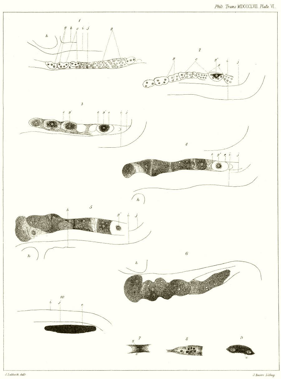
EXPLANATION OF THE PLATES.
PLATE VI.
Fig. 1. Ovary of Daphnia Schœfferi, shortly after the deposition of agamic eggs in the receptacle.
Fig. 2. The same, a few hours later.
* Loc. cit.
† Ann. and Mag. of Nat. Hist. 1852.
‡ Wahre Parthenogenesis, &c.
[page] 100
Fig. 3. Subsequent changes in the same ovary.
Fig. 4. Subsequent changes in the same ovary.
Fig. 5. Subsequent changes in the same ovary.
Fig. 6. Subsequent changes in the same ovary.
Fig. 7. Early stage of the ephippial egg.
Fig. 8. The posterior part of the ovary, showing the little balls into which the embryonic ephippial egg often breaks up.
Fig. 9. Ephippial egg more developed. It has become so opake that the germinal vesicle cannot be seen.
Fig. 10. Ephippial egg nearly ready to be laid.
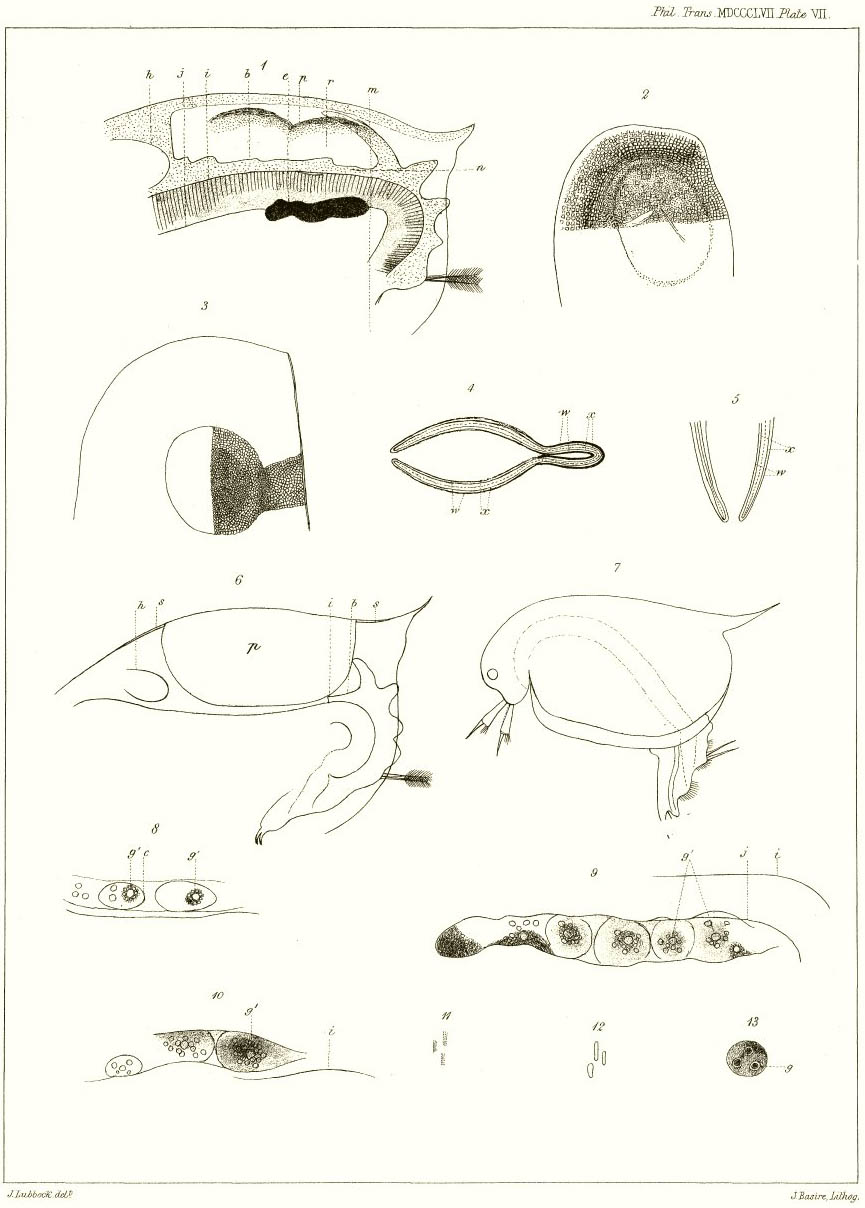
PLATE VII.
Fig. 1. Part of the back of a specimen of D. Schœfferi, ready to lay an ephippial egg.
Fig. 2. Half of the inner valve of an ephippium, only in part finished.
Fig. 3. Half of the outer valve of an ephippium, only in part finished.
Fig. 4. Diagram of a section of the carapace along the line m. The body is omitted.
Fig. 5. Diagram of a section of the carapace along the line n. The body is omitted.
Fig. 6. Part of a female Daphnia Schœfferi, showing the eghippium in outline.
Fig. 7. Outline of a male Schœfferi, to show the male generative organs (q).
Fig. 8. Part of an ovary of a D. Schœfferi, to show agamic eggs just beginning.
Fig. 9. Part of an ovary of a D. Schœfferi, to show agamic eggs in the process of formation, each round a distinct germinal vesicle (g′).
Fig. 10. Ditto of another specimen.
Fig. 11. Contents of ephippial egg rolled together.
Fig. 12. Contents of agamic egg rolled together.
Fig. 13. An ovarian mass, containing three ovarian cells, any of which may become a germinal vesicle.
The letters refer to the same parts in all the figures:—
b. Line of the back of the body.
c. Ovarian masses.
e. Early stages of the ephippial egg.
g. Ovarian cells, some of which become germinal vesicles, g′.
h. Heart.
i. Upper line of intestine.
j. Lower line of intestine.
p. Ephippium.
q. Penis.
r. Receptacle or space between the back and the shell.
w. Outer and inner layers of old carapace.
w′. Outer and inner layers of old carapace modified to form the ephippium.
x. Outer and inner layers of new carapace.
[Plate VI]
[Plate VII]