[page] 124
2. Notes on Entozoa. Part II. By T. SPENCER CORBOLD, M.D., F.R.S., F.L.S., Lecturer on Parasites at the Middlesex Hospital Medical College.
[Received January 2, 1874.]
(Plate XVIII.)
Whilst engaged in writing the concluding portion of my first set of "notes," I received for examination a nematode parasite, some brief account of which will appropriately commence the present series.
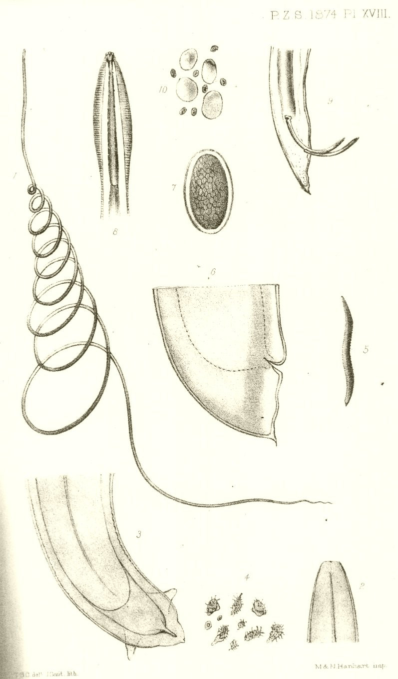
4. FILARIA GRACILIS, Rud. (Plate XVIII. figs. 1-4.)
On the 20th of August, 1873, Mr. Samuel Smith, M.R.C.S., of Clifton, transmitted an example of this entozoon, with a request that I would identify it. Finding that the specimen was a male, and unaware that the males of this species ever attained a length of 20 inches, I at first supposed that we had to deal with a new form. However, on subsequently analyzing its characters, I became satisfied that the worm was really only an unusually fine male F. gracilis. The frequency of the occurrence of this nematode in the abdominal cavity and other parts of the trunk of Monkeys is a matter of common observation. I remounted no less than four preparations, representing numerous examples of this Worm, for the Museum of the Royal College of Surgeons. Some of the Hunterian specimens were originally obtained by Professor Owen from the cavity of the pleura of a Capuchin Monkey, others having been removed by him from the thorax of an Orang-outang. From Mr. Smith I have learnt that the present example formed one of a group of five Worms, all of which were found lying between the folds of the omentum of a Spider Monkey (Ateles). My informant also remarks that one of the Worms was enclosed in a "false sac, formed by a twisting of that portion of the serous membrane which is immediately connected with the inferior curvature of the stomach." It further appears that the example in question was the smallest of the five, although I found it to measure upwards of twenty inches without any stretching. Whilst the Vienna helminthologist, Diesing, only allowed an extreme length of four inches for the male worm, the French authority, Dujardin, stated that specimens had been reported up to a length of 12½ inches. Females have been recorded as reaching a a length of 5 feet.
Not being acquainted with any satisfactory representation of this Entozoon, I have thrown the parasite into a series of folds so as to enable me to display its full length and general appearance (Plate XVIII. fig. 1). I have also added an enlarged and accurate outline representation of the head and neck (fig. 2). The description of the Worm by Dujardin leaves little or nothing to be desired. Speaking of the tail, he observes that the extremity is furnished with two or three papillæ, serially disposed in front of or above the point. I examined
[page] 125
these minute prominences carefully (fig. 3). There were two short conical papillæ placed within about the 1/800 of an inch from the actual extremity, the point itself being furnished with an excessively minute prominence, whose base scarcely exceeded the 1/10000 of an inch in diameter. There was a very distinct appearance of a centrally placed duct (which I regarded as the tubular extension of a large caudal gland), the end of which had apparently become detached from the interior of the minute terminal papilla. As already remarked by Dujardin, the lateral lines of the body are brown-coloured and very conspicuous. I was particularly struck with the remarkable distinctness of the contents of the seminal tubes, whose separate particles could readily be seen through the thick integuments. Unwilling to injure the specimen, which I afterwards returned to Mr. Smith, I merely inserted the point of a fine needle into the main channel, and thus obtained a large quantity of the spermatozoa. These small particles, notwithstanding their long immersion in strong spirit, presented a tolerably characteristic appearance—the larger and fully formed corpuscles giving a long diameter of 1/1400 of an inch (fig. 4). I may add that several of the corpuscles displayed, more or less perfectly, the well-known flask-shaped envelopes so often described in connexion with this group of parasites.
5. SPIROPTERA TURGIDA, Duj. (Plate XVIII. fig. 5.)
On the 15th of April, 1873, I examined the contents of a small phial in which were two Worms that I had long previously received from Dr. Murie. They were sent to me during the time of his official connexion with the Society's Menagerie. The smaller parasite, as was stated on a label, came from the stomach of an Opossum (Didelphys azaræ). The worm was evidently a female, but, unfortunately, not in a satisfactory state of preservation. It measured more than an inch in length by 1/10" in breadth. The accompanying figure may be useful (fig. 5); but the minute characters were mostly either lost or obscured. The mouth was round, and certainly furnished with several minute teeth, the number of which could not be accurately ascertained.
6. ASCARIS CUSPIDATA, T.S.C. (Plate XVIII. fig. 6.)
The larger of the two Worms above-mentioned appears to be new to science. Dr. Muria labels it as having been obtained from the stomach of a Green Monkey. I have little doubt that his record refers to one of the Monas (Cercopithecus). The Worm is a true Ascaris, and, although a male, measures fully 3¾ inches from head to tail. Owing to imperfect preservation, the spicules have been lost. The three oral lobes are particularly prominent. The caudal extremity is furnished with a very fine spine, or cusp, formed by an extension of the epidermis (fig. 6). This minute cusp curves backwards, and measures only 1/1000 inch in length by the 1/3000 inch in breadth at the narrowest part. The anal aperture is placed at a distance of 1/50 inch from the extremity of the tail. The eggs have a long diameter of about the 1/300 of an inch.
[page] 126
7. ASCARIS MACULOSA (Rud.). (Plate XVIII. figs. 7-10.)
On the 9th of October, 1873, I received a letter from Dr. J. Alexander Macdonald, of Woburn, Bedfordshire, stating that he had forwarded to me a pigeon which had been found dead on the previous morning. It seems that the owner of the bird had erected a large pigeon-house, and had imported a number of Antwerp Sweeter, these birds all continuing in a perfect state of health until about a week before the above-mentioned date, when, to use Dr. Macdonald's words, "first one and then another was attacked, and so on, until four or five of the pigeons had died after a few hours' illness." The suddenness of these attacks not unnaturally suggested poisoning; and, accordingly, says my informant, the owner "had the curiosity to open one of the birds, when, to his astonishment, he found the intestines stuffed with worms."
Under these circumstances I was invited to make an accurate inspection of the pigeon forwarded to me, likewise to report the results of my examination, and to suggest any remedial or prophylactic measures which might be likely to prove useful.
Two days later I received a letter from Dr. Macdonald stating that several others of the flock had died, and it further appeared to him probable that the daily list of sick and dying would continue to increase. On the 14th of the month my informant reported that three more of the birds were dead ; but this mortality still left twenty-five birds in the owner's possessson, some of which were affected. Under these circumstances I lost no time in forwarding a full report of the facts observed, together with the recommendation that a few grains of santonine should be mixed with the food. Speaking of the birds seen on the 14th October, Dr. Macdonald says that "one which appeared in a hopeless state Was at once treated (by the owner) to two grains of santonine;" and when my informant saw the bird in the afternoon of the same day "it had so far recovered as to be hopping about and picking up food." On the 4th of the following November the same correspondent obligingly informed me that the epidemic had been "at last mastered." It seems that altogether twelve birds had perished, the remainder now appearing perfectly healthy.
It is not stated whether the final and satisfactory result appeared to be due to the administration of the remedies I had recommended; but, in any case, the cessation of the disorder following so close upon the employment of santonine is worthy of being noticed. I had almost hoped that my report, in the interests of science, would be published; but, so far as I am aware, such has not been the case. In regard, however, to the dissection (upon which that report was mainly based), I have felt sure that the scientific and practical data it supplied were of sufficient interest to be placed on permanent record. The necessary dissections and microscopic examinations were made on the 9th and 10th of October, whilst the bird was perfectly fresh. The blood, muscles, and cellular tissues, and every organ of the body, apart from the digestive apparatus, were found to be thoroughly healthy; and it was only when the alimentary canal
[page] 127
had been laid open that I found any visible traces of parasitic disease. From the lower opening of the crop downwards to the termination of the small intestine the canal was more or less crowded with nematode entozoa, all of them being referable to the above-named species. In spite of this state of things, and notwithstanding, also, that the small intestine was inflamed throughout (showing several large ulcerated patches, which here and there measured fully an inch in length), the body of the pigeon exhibited no traces of emaciation. From this it was evident that the parasites had developed rapidly, and that the malady had a correspondingly rapid formation. The local distribution of the parasites themselves was especially noteworthy. One specimen, two inches long, extended from the crop to the proventriculus. The cavity of this latter organ and also that of the gizzard were crammed with worms, which completely blocked the passage between the two. Three of the worms had also placed themselves within the pyloric opening, their bodies partly lodging within the upper part of the duodenum. The duodenum itself was crowded with these Ascarides; but their numbers somewhat decreased towards the lower folds of the small intestine. I removed 36 worms from the œsophagus, proventriculus, and stomach, besides 166 others from the intestinal canal, thus obtaining a total of no less than 202 nematodes from this solitary avian bearer. Considering the comparatively large size of these entozoa, this high degree of infection must be pronounced remarkable. The largest female worms measured 2½ inches in length. One of the most interesting facts—serving to exemplify a well-known habit of lumbricoid worms generally—consisted ire the circumstance that two of the parasites had succeeded in perforating the horny lining membrane of the gizzard. The injuries had evidently been accomplished during the life of the host, since the walls of the gizzard were inflamed opposite the perforations made by the heads of the parasites. There was a little half-digested and green food within the stomach, the débris of which, when placed under the microscope, showed several characteristic nematode ova. There were no free embryos anywhere discernible; neither had the development of the freed eggs proceeded beyond the coarsely granular stage of yolk-segmentation. Free eggs were also found both in the small and large intestine. The eggs measured about 1/360 inch by 1/700 inch in diameter (fig. 7). An admirable description of the adult parasite has been given by Dujardin; but since the published figures of Rudolphi, Goeze, and Bremser are incomplete, I have thought it desirable to supplement them with others. Thus fig. 8 gives a magnified view of the head of a small female Ascaris maculosa, and shows especially the crenulations or transverse striæ on the alæ or lateral membranous appendages. These are veritable annulations, or fine circular striæ, and not merely contractions of the integument as some have supposed. Dujardin speaks of the alæ as scarcely visible; but I always found them more or less marked and semielliptic in shape, as Rudolphi originally described them. Another sketch (fig. 9) shows an enlarged view of the tail of the male; and, on the whole, this representation agrees
[page] 128
very well with Bremser's figure of the same part. The arcuate spicules, however, are not so sharp at their tips as his illustration implies, and they are certainly more uniform in thickness. Dujardin remarks that Rudolphi has represented the spicules as being straight, whereas be himself always found them curved. Rudolphi, however, was scarcely in error, since I have repeatedly noticed that these arcuate organs are very nearly straight in their perfectly retracted condition. Like Dujardin (and without having previously consulted his descriptions), I was particularly struck with the appearances presented by certain large perivisceral corpuscles, the presence of which originally suggested the specific name of the worm. Dujardin very appropriately calls them corpuscules orbiculaires diaphanes, but compares them, somewhat unfortunately, to little acephalocysts. These bodies, as he says, are many times larger than the ova. For my own part, I believe they are nutritive in character, and, like the fluid in which they float, are, I suspect, chemically comparable to the juice of flesh. At all events, Dr. Marcet has proved that the perivisceral fluid of the large lumbricoid worm of the horse (Ascaris megalocephala) partakes of this character; and it is no uncommon thing to notice similar corpuscles in the bodies of other nematode worms. Dujardin himself refers to similar bodies in an Ascaris from the Perroquet. I have purposely represented a few of the eggs along with the nutritive corpuscles, side by side, in order to show their relative sizes (fig. 10).
Notwithstanding the facts thus set forth in connexion with the parasitic epidemic affecting the Antwerp Smerles, the entozoa in question do not appear to be very common. Dujardin has remarked that Heister, at Rostok, and Gebauer, at Breslau, found this parasite abundant at the beginning of the 18th century; but, according to examinations conducted at Vienna, the worm was found in the Common Pigeon in only 11 instances out of 245, and thrice only in 38 examples of the Ring-Dove; moreover the examination of 87 other pigeons and doves of different species yielded entirely negative results. These data are of high practical interest, and they serve to throw light upon questions of epidemiology. I may add that the Dublin helminthologist, Bellingham, long ago noticed the occurrence of this parasite in Ireland.
EXPLANATION OF PLATE XVIII.
Fig. 1. Filaria gracilis: male, nat. size.
2. The same: head and neck, enlarged 40 diam.
3. The same: tail, mag. 70 diam.
4. The same: spermatozoa, mag. 350 diam.
5. Spiroptera turgida: nat. size.
6. Ascaris cuspidata: tail, enlarged.
7. Ascaris maculosa: egg, mag. 330 diam.
8. The same: head of female, mag. 20 diam.
9. The same: tail of male, mag. 35 diam.
10. The same: eggs and nutritive corpuscles, enlarged.
[Plate XVIII]