[page] 365
XVIII. Some Observations on the Structure and Functions of tubular and cellular Polypi, and of Ascidiæ. By JOSEPH JACKSON LISTER, Esq. F.R.S.
Received January 1,—Read March 6 and 13, 1834.
THE more obscure functions of vitality are of such difficult investigation, and possess at the same time so high an interest, that any one contributing, in however small a degree, to increase our information regarding them, may hope to meet with indulgence.
This consideration encourages me to submit to the Royal Society some observations made during a few weeks spent at Dover and Brighton in the autumn of 1832, and the last summer. I was led to engage in them from having two years before noticed the existence of currents within the tubular stem of a species of Sertularia; and their investigation has led me on to additional particulars relating to that family of zoophytes, and other compound animals more or less resembling them, some of which I am willing to hope may be new in physiology.
The facts being only such as presented themselves during a limited stay on the coast, and in part indeed requiring further observations to ascertain their true bearing, the form of the original memoranda is often retained, as being probably the most satisfactory. Though too circumscribed and incomplete to form a ground for new arrangements or theories, they will at least show that the field from which they are culled is hitherto but partially explored, and may perhaps awaken the attention of inquirers more favourably circumstanced.
Of the notices regarding tubular polypi, one on Tubularia indivisa relates principally to a peculiar circulation seen within it, and to some circumstances attending its growth, absorption, and decay. Those which follow on Sertulariæ describe internal currents of a different kind, a more full observation of growth and absorption, with a case of the development of ova, and some other particulars of this family.
An account is next given of a minute Ascidia possessing a character, not I believe before observed in that tribe, of distinct individuals connected by a branching stem and a common circulation, and bearing in these and other points some resemblance to the Sertulariæ. The organization and functions of this Ascidia, and of a Polyclinum allied to it, are stated the more in detail, as the anatomical descriptions of the Tunicata by CUVIER, SAVIGNY, and MACLEAY appear to be derived almost wholly from dissection.
There are at the conclusion remarks on the natural character of some Flustræ and other cellular polypi, which tend to confirm the opinion that they are of a family
MDCCCXXXIV. 3 B
[page] 366
much nearer to the Ascidiæ than to the Tubulariæ or Sertulariæ with which some of them have been hitherto associated.
It may deserve to be mentioned, that as the species of animals of one type diminish in size, the delicacy of some of the remoter details of their structure does not increase in the same proportion; but the aggregation of the component parts in the smaller species becomes instead more simple. From this cause, as well as from their transparency, most of the objects examined offered peculiar advantages for inspecting their organization while living and in freedom. They were placed in a glass trough with parallel sides, before my achromatic microscope directed horizontally, and the sea water was often changed. Near the end of the observations this was done by two siphons, one of which constantly admitted a fresh supply, while the other carried off the excess; a mode which, had it been earlier adopted, might have rendered some of the results more satisfactory; for the great difficulty, next to procuring a variety of specimens, was to retain them in vigour.
With the exception of two species that grew above the line of low water at spring tides, those obtained were only what the waves threw up; for repeated endeavours to get them, through fishermen, from rocks at sea or from oyster-grounds were unsuccessful.
The drawings in illustration were traced by a camera lucida slid over the eyepiece of the microscope. The facility with which correct graphic records and measurements may be obtained by means of that instrument with a little practice, induces me to recommed its use to other observers. The linear enlargement is marked to each figure.
TUBULAR POLYPI.
TUBULARIA.
The specimen of T. indivisa figured Plate VIII. fig. 1., was one among a broken mass of tubes, most of them larger than itself, which was found at Dover, October 1832, freshly cast ashore, and was kept several days, on another account, before the polypus terminating it was observed. It is a very small example of the species to which it is considered to belong. The arms had no ciliæ.
When magnified about one hundred times, a current of particles was seen within the tube, that strikingly resembled, in its steady continued flow, the circulation in plants of the genus Chara. The general course of the stream was parallel to the slightly spiral lines of irregular spots on the tube, and in the directions marked by the arrows. On the greater part of the side first viewed (that shown in the drawing), it set as from the polypus; but on reversing the glass trough so as to show the other side, the flow was there towards the polypus; each current thus occupying half of the circumference. The particles had no dancing motion among themselves like those which will be hereafter mentioned in Sertularia, but floated evenly on at a uniform rate. They were various in size; some very small, others apparently aggrega-
[page] 367
tions of smaller ones; a few were nearly globular, but in general they were of no regular form. The tube had between the lines of more conspicuous spots a granulated appearance, and beneath this the currents ran; but I could not, by altering the focus of the microscope, detect the opposite current on the further side, as may be done in Chara, owing to the interposition, in most parts, of a grumous matter. Some of the larger particles of this were like those in circulation, and by close attention might be seen slowly to change their relative positions.
At the nodous parts c d e were slight vortices in the current: at c near the end of the tube it came over from the opposite side, and I could not at this time succeed in detecting the passage of any particles between the tube and the stomach of the polypus.
Between the stomach b, and the mouth a, a remarkable action went on, wholly different from that in the tube. The mouth became swollen by a flow into it from the stomach, to the shape shown at a 3: this flow continued for about a minute. The contents of the mouth were then squeezed back into the stomach, which expanded as the mouth contracted (a b). During this reflux the connecting orifice was seen distinctly open, and it continued so on the return of the flow to the mouth (a 2), till the stomach became nearly emptied. The orifice then closed gradually, preparatory to the effort of forcing the fluid back to the stomach. The intervals between that act were very evenly eighty seconds.
Two currents were continually going on, both in the mouth and stomach, one flowing always down the sides in the direction a b, and an opposite one in the axis; except that the latter was suspended at the time of the close contraction of the stomach and of the discharge from the mouth into it.
The creature was observed at intervals throughout the day from ten in the morning; its appearance continuing much the same. The front arms sometimes spread themselves a little, and at one time a cloud of particles hung before them in the water like those within the mouth, and which seemed to have been recently ejected. Though the swelling and contracting continued, no motion was seen in the ejected particles; proving that in the ordinary course of this action the mouth was not opened.
The next morning the polypus was found much altered (a 4); the hinder arms, or those of the neck, were shrunk to an indistinct mass, appearing to be partly absorbed and partly thrown off, and a stream of particles was drifting away from them. The least imperfect of these arms had a rapid action going on in its granular substance, which ended in a current within it towards its root. The terminating arms appeared as perfect as the day before.
Rudiments of the horny shell had extended to the neck from f, its end when it was first observed. The currents in the intermediate space b were now plainly connected with those in the tube and had lost their former even course, the particles flowing about irregularly as the division between b and c became by degrees broken away. The currents in the tube were unaltered.
3 B 2
[page] 368
Later in the day the imperfect new piece of shell was folded in, the stomach b had disappeared and the end of the soft part within the tube was shrunk to a conical form, all dilatation and contraction (of which there were some remains in the morning) having ceased; another discharge had taken place from the mouth, and a considerable one from the neck, when the tube acquired a defined end (a 5); and between the shell and the conical soft part a vacant space had been gradually left. Into this was now admitted from the neck some granulated fluid matter, which flowed over the conical surface, and after some agitation of the particles, formed a rounded covering to it; semitransparent at first, but soon becoming more opake, so as not to be distinguished from what it covered (a 6). The circulating particles of the tube now flowed into the end and returned from it, as at a septum of Chara.
Afterwards rapid action of large granulations and stillness alternating took place within the neck; and in the evening the polypus was completely separated from the soft matter of the tube, and dead; its substance breaking away in a stream of disengaged particles. The following morning some slimy matter about the end of the tube was all that remained of it; but the circulation within the tube continued as before.
The specimen was now immersed in spring water. The first effect was, instead of the two currents, one flow, towards the end of the tube, of roundish particles, some of which escaped into the water: after this had continued a short time, the whole internal contents of the tube began to move forwards together, and became protruded at the end like thin paste; then for a while a clear current with few particles in it flowed back between the shell and the moving mass, and showed the shell to be transparent, and that the mottledness and granulations, as well as the irregular lines of spots which were before seen in it, were wholly belonging to the soft matter; another circumstance analogous to what is met with in Chara.
This single observation on Tubularia was prolonged in the hope that the destruction of the hinder arms might have proved to be part of a process of lengthening the tube. Some of the later appearances were evidently the effect of disease, and the action between the mouth and stomach during the first day seemed unnaturally violent; yet the currents there will be seen to bear a resemblance to those which accompanied vigorous growth in Sertularia. The instance of deposition recorded stands at present by itself; that of absorption will be fully corroborated. The circulation in the tube exhibits a character hitherto, I believe, only observed in the vegetable kingdom.
SERTULARIÆ.
Each of the divisions Tubulariæ and Sertulariæ, as adopted by CUVIER, includes widely differing genera, while they separate others that are closely allied. Some which I think should be placed among cellular polypi, will be noticed hereafter;
[page] 369
others I have not seen; but Plumularia, Sertularia, and Campanularia belong to one very beautiful natural family, for which the old name of Sertulariæ may perhaps be well retained, and these are the subject of the observations under the present head. The specimens figured include some principal variations in the form and position of the cells.
Sertularia pluma, Plate VIII. fig. 2. (ELLIS, pl. VII. b B. Plumularia pluma, LAMARCK), was found at Dover, October 1832, on the ropes of Fucus siliquosus. It consisted of a minute horny tube, creeping along and round the Fucus with frequent anastomosis, (a usual property of the Sertulariæ,) and sending out offsets at short intervals. These had each a main stem, feathered with jointed branches directed to each side alternately in close succession: every joint was elaborately formed of thin transparent shell, of which one part continued the branch, and the rest formed a cell for a polypus; all the cells on a side branch taking one direction. Each plume might comprise from 400 to 500 polypi. It was neglected to be drawn till the polypi were shrunk: when living, their arms spread widely, their body scarcely projecting beyond the edge of the shell.
All the polypi were connected together by a soft matter, of a pulpy or finely granulated appearance, which was a continuation of their substance, and extended throughout the interior of the branches, the stem, and the creeping tube or root. When a magnifying power of 100, and still more clearly when one of 300, was used, a current of particles, various in size and of irregular form, was observed running along the axis of this soft matter. It flowed in one channel, alternately backwards and forwards, through the main stem and lateral branches of a plume, and through the root as far as the opacity admitted of its being traced: sometimes it was seen to continue into the cells. The stream was throughout in one direction at one time: it might be compared to the running of sand in an hour-glass, and was sometimes so rapid in mid-tide that the particles were hardly distinguishable; but it became much slower when near the change. Sometimes it returned almost without a pause; but at other times it was quiet for a while, or the particles took a confused whirling motion for a few seconds, the current afterwards appearing to set the stronger for this suspension. The whirling or starting motion took place sometimes at one sometimes at another part of the stem and branches during the intervals of the currents. Five ebbs and five flows occupied fifteen minutes and a half, the same average time being spent in the ebb as in the flow. The longest continued stream was two minutes and a quarter; the longest suspension, half a minute.
When the connexion of a plume with the root was interrupted by bending its stem, the stream running down the middle was observed to continue its flow up one of the lower and stronger lateral branches, and then to return down that branch and up the main stem, the course of the current in most of the other side branches being still the same as in the middle one.
On a stem being cut off below the commencement of the side branches, a few
[page] 370
seconds passed before anything exuded from the stump. A small stream of particles then issued, soon succeeded by a flow of viscid matter: this stopped a while, and then went on again, but ceased altogether in about five minutes. It hung like honey about the end, and on its gradually clearing away, the wound appeared healed. The successive flow, slight agitation, and reflux of particles, continued in the stump as before the separation. The alternate currents in the axis of the soft matter were seen in all the Sertulariæ that were examined, and appear to be an essential character of this family. Further particulars regarding them will be noticed as they were observed in other species.
I am not aware that any writer of our own country has mentioned the existence of these currents, unless it be Dr. FLEMING, in an account of Sertularia gelatinosa given in the Edinburgh Philosophical Journal, 1820*; but his description, if accurate, must refer to a different action. Some of the continental works which were the most obvious to be consulted are silent regarding them; but on further search, in CAVOLINI'S "Memorie per servire alla Storia de Polipi Marini," published at Naples in 1785, the general character of the motion was found very clearly and explicitly given.
It is extraordinary that so singular a phenomenon as the flow and reflux of the circulation in one and the same channel, announced evidently by a careful and able investigator, should have remained, as it appears to have done, almost unnoticed for nearly half a century. I am glad to corroborate these observations by a quotation from his treatise, which serves also to explain some of his opinions†.
* "When in an active state, I have observed the water taken in at the mouth descend, for the space of several seconds, through the gelatinous parenchyma of the body and footstalk, and again return to be ejected. The fluid thus circulating did not seem to move in a body through tubular vessels, but to be divided into minute globules, which permeated a cellular structure."—Edinburgh Philosophical Journal, vol. ii. p. 85.
† "Un fenomeno assai singolare nell' economia delle Sertolare è un movimento che si osserva nell' interiore del corpo come in un proprio tubo. L' esteriore corneo invoglio, ordinariamente transparente, chiude e veste il corpo molle dell'animale, il quale corpo si vede essere formato come di un amasso granelloso. In mezzo di questo corpo per una linea alungo si vede che una simile granellatura venga transportata con moto vorticoso di un fluido che non si arriva a distinguere: mercè di questa agitazione si vede che quelle briciolette di materia ora vengono portate in giro, ora in una corrente salgono in sopre or discendono; e questo fenomeno accade così nel tronco principale che nei rami fino a toccare gli organi polipiformi e dura ciò finchè vive la sertolara anchorchè i suoi organi siano strettemente ritirati."—p. 121.
"Questo corrente si vede estendersi per tutto il tratto del corpo di esso polipo (Sertularia Dichotoma) e fino alla testa dei polloni che si sviluppano nel mezzo dei divisati calici; si arresta poi nel piede di questo quando si sono sviluppati in organi polipiformi….. Questo canale altro non può essere che il cuore; in fatti quando i polloni si sviluppano in organi polipiformi siccome questi devono predare e digerire il cibo non può il cuore più apartenere a loro per che devono serbare un organo ministro del cuore. Ma è cosa degna di considerazione che per questo cuore discendano briciolette medesime che sembrano entrare nella composizione del corpo dell'animale…. e se potrebbe dire che queste briciolette di materia son quelle che ricevuta avendo certa alterazione dall' azione del cuore, si animalizino, cioè passino nella formazione del corpo stesso dell' animale. E queste briciolette dovranno necessariamente al cuore venire dai ventrigli, i quali posti nel fondo degli organi polipiformi triturano gli animaluzzi infusorj che dalli tentacoli sono acchiappati."—p. 197.
[page] 371
CAVOLINI saw the currents in all the Sertulariæ observed by him, but he did not detect their continuation into the stomach of the expanded polypi; a circumstance equally belonging to every species that I have met with. Had he done so, he must, it would seem, either have included the stomachs, with the branches and the root, under the name of heart, or probably would have relinquished a term which, on several accounts, appears to be misapplied. His not having perceived the stream of communication may be accounted for by its being generally much smaller and less conspicuous in a short space near the base of the cells, as well as less regular in its periods, than that which runs along the stems and footstalks.
Sertularia pumila, Plate VIII. fig. 3. (ELLIS, pl. v. a, A.), was gathered growing above low water mark at Dover, October 1832. Each of its cells is divided near its base by a partition a, on which the polypus is fixed, and which may be seen to be pierced with an oval hole b, if a section of the shell is made a little above or below it. In the complicated cell of S. pluma, last described, this perforated septum was not detected; but I believe it to exist throughout the family. Sertularia abietina has the aperture long and narrow. In Campanularia it seems to be round, and in the middle of the septum, which in that genus is very conspicuous.
The connexion between the stomach and the stem, by the aperture, takes an angular course in the present species, and could not always be perceived. One polypus had within its mouth revolution of darkish substances, which after a while it disgorged; in the stomach of the same there was irregular motion of particles, and an action seemed to be going on between the two; the upper part of the neck sometimes swelling, and the cavity of the mouth extending down that portion in a tubular form. Within other expanded polypi, no motion could be detected.
In the substance of the necks of the polypi, transverse lines were visible, bearing a resemblance to those characteristic of voluntary muscle in the higher animals. I have observed the same appearance on a band forming the edge of the bag of a Lucernaria, and also very distinctly in the axis of its numerous knobbed tentacula.
Sertularia setacea, Plate VIII. fig. 4. (ELLIS, pl. XXXVIII. 4 D. Plumularia setacea, LAMARCK,), was found at Brighton on flag, with which the shore was strewn after a storm, July 1833. It was distinguished by its subconical cell, so short as commonly to shelter only a part of the stomach, and by its spinous ovaries, differing from those in ELLIS'S figure, and mostly sessile on the creeping root. The ova within were opake and yellow. Its polypi had from sixteen to nineteen arms, and when they were full-blown it was an object of remarkable beauty. From its transparency, and the smaller number of its moving particles, their individual quivering motions and the course of its currents were more conspicuous than in the former two species. The stream sometimes extended only to the pulp below the septum, and sometimes mounted into the stomach; and in whichever part it terminated, agitation took place there on the ceasing of the upward flow. The soft part within the branches,
[page] 372
which adhered generally to one side of the tube, had the look of a slimy matter, inclining to granular, and held together by greater tenacity at its outside. Nothing like muscular contraction was seen in the pulp of this or any other species.
As a little globular animalcule was driving rapidly past one of the expanded polypi, it instantaneously seized it, and brought it to its mouth by contracting its arms. They gradually opened again, except one, that remained a while doubled, with its end on the animalcule. The mouth indistinctly seemed filled with hairs or tentacula, that closed over the prey; and after a few seconds it was carried slowly down, in the manner of the Actiniæ, the mouth contracting and the neck enlarging, into the stomach: here it was uncertainly seen, and soon disappeared. Agitation of particles in the stomach followed the swallowing, and then the currents between the stomach and the branch went on again as usual.
One polypus was supported on a stalk that, probably owing to injury, was entirely empty of pulp; yet it was still open and vigorous.
The minute and delicate species, Plate VIII. fig. 5, found at Brighton July 1833, resembled Campanularia in having its cells placed on footstalks, and in the branching of its stem, but was differently jointed, and the cells and polypi were nearer to Sertularia pluma. The polypi had sixteen arms. The currents, which generally extended into the stomach of each, set more strongly into the side appendage, which they all possessed, ending in two small ears, and looking like a continuation of the footstalk. All the shell of the cells, when dry, gave the colours of thin plates, owing to its extreme tenuity.
The only ovary, a, in the specimen was enormously large compared with the cells, depressed at the end, and transparent. It had an opake ovate substance within, of a dirty yellow colour, that was connected with the base and with the extremity of the ovary by a column of soft matter running nearly in the axis. Within this, between the base and the ovate body, currents were seen like those in the stem, and sometimes a motion of particles was observed in the adhesion to the shell at b. No separate ova, like those to be noticed in Campanularia, were visible.
Several species of Campanulariæ that were observed agreed very nearly in the form of their bell-shaped cells; with a distinct septum, and a thin column of soft matter between it and the base of the cell. The branches also of all had annular indentations round them, more or less numerous, which form a simple and beautiful provision for giving flexibility combined with strength.
The specimen figured in Plate IX., or that in Plate X. fig. 1, may equally serve, I believe, as an example of Sertularia dichotoma, (taking that to be the name of ELLIS'S species, pl. XII. a, c, C. and XXXVIII. 3, B.); but the former of the two was much the stouter in its growth. Its cells were from ·018 to ·02 inch in length, and the arms of its polypi commonly thirty; the other had about twenty-six arms and cells not exceeding ·014 inch.
[page break]

[page] 373
The young branch, Plate IX., furnished an instance of the progressive production of a polypus; for having been drawn (August 19, 1833,) on account of the singular form of the cell b, the sprout c was found to have lengthened during the tracing, and this led me to watch its growth at intervals during that and the following day. About five hours were spent in the completion of the footstalk, and twenty-seven more in that of a cell and its polypus.
The branch sprang from a creeping tube that had been recently broken at a little distance beyond. This, like the pruning of a tree, may have accelerated the growth; and from the same cause as was supposed, the internal currents were very irregular. Dancing of particles sometimes took place along the whole stem, sometimes at parts of it only. The set of the stream was sometimes up or down a b and c altogether, sometimes out of a into c, or the contrary. Before a current in the stem became strong, some particles would for a while appear to struggle on against it. The sprout c, which looked like a continuation of the stem, was at first quite full of soft matter, and the motion of the currents in it was inconsiderable; but they increased so as soon to become greater by far there than in any other place, and its soft part was then dilated and contracted alternately, except at its end, which was always full. The time of the stream's running into c was, on an average of seven alternations, six minutes; of the reflux four minutes. The longest time of the former was eight minutes and a half, the shortest of the latter two minutes. In about two hours the sprout had assumed the appearance c 2. Particles of larger size were now seen, maintaining a continual circulation within, except that all was still for a short time when the pulp was most shrunk: when it was dilated they had great agitation. Their flow was always downwards along the axis, and upwards along the sides of a defined cavity, being in a direction opposite to that observed in the polypus of Tubularia; and the manner of the flow, and the abundance of fluid, more resembled the circulation of Chara than anything I had before seen in Sertularia; but still the dancing motion distinguished them. Some of the particles were nearly round, and transparent, other larger ones seemed like masses of the pulp brought into circulation.
The part of the branch towards the end was more opake than the rest: slight currents were occasionally seen in the axis there at long intervals; and the outer portion of this part, which was in the act of growth, had not a granulated appearance, but was marked with radiating lines nearly parallel to each other in front, and more diverging on the sides, so as to make always a considerable angle with the surface; and among them no current or motion was seen.
In six hours more the branch had the shape of c 3, the rudiments of a cell f appearing at the end: its commencement was about three hours previously. The peculiar circulation down the axis and up the sides was now only during the latter part of the influx; the dilatations were lessened, and a part of the shell at d was never filled; a separate agitation had begun to show itself at e, and a faint one at f.
MDCCCXXXIV. 3 c
[page] 374
After being left for a night, the incomplete cell had grown, in twelve hours from its beginning, to the appearance c 4. The soft matter of the branch was now wholly detached from the shell on the left side, but lay close along it on the right: the currents in that part were become scarcely perceptible, very slight dilatation and shrinking being sometimes the only indications of them. The two tracings c 5 give alternations in the soft matter of the cell three hours later: the striæ indicative of the forming process, smaller than those seen during the growth of the branch, still remained near the end: when viewed as an opake object, the part f was a brownish orange, and g milky. In a short time rudiments of arms appeared; and c 6 represents them when the cell seemed completed after sixteen hours' growth: its end, however, was closed, and the polypus, by slow motions backwards, forwards, and sidewise, was releasing the yet imperfect arms from their adhesion to the side. The tracings c 7 and c 8 show their continued progress.
At twenty-six hours from the commencement of the cell the arms, apparently fully formed and folded over each other, had been pressing against the end of the shell, which seemed still to inclose them but had something of a ragged look, when at length several were slowly raised beyond the cell. On their being drawn in again, a little transparent film was seen projecting, the remains, as appeared, of what had covered the end. The arms were then again protruded as at c 9. The stomach enlarged and contracted, but seemed coated with an orange-coloured matter in irregular masses.
In one hour more the branch was terminated by a fine perfect polypus, fully expanded, with twenty-eight arms, which was discharging at the mouth the opake orange-coloured matter that had lined the stomach.
The mouth, when open, varied from the form of a saucer to that of an upright cup; no hairs were detected within it.
The pulp in the stalk had now many fresh adhesions to the shell on the left (c 9), and must therefore have swollen since the tracing c 4, and contracted again.
For some time after this the polypi of b and c continued expanded and, to appearance, healthy; but that of a, which on the first day gave proof of its vigour by seizing and swallowing an animalcule, shrivelled greatly during the night, and was found in the morning to be evidently in the course of absorption. A downward flow from it of particles that were mostly small and a few orange-coloured, took place every eight or ten minutes, continuing for three or four: the influx in the intervals was scarcely or not at all visible. The progress and completion of this absorption are shown in the tracings a 2, a 3, a 4, a 5.
Another Campanularia, resembling the above, after being kept some days, though fresh sea-water was often supplied gave the following symptoms of decay. The polypi which remained were all contracted, with great agitation of particles in their interior, and increase of the currents. Next, the continuity of the soft matter which had connected them with the stem became broken, and no further motion was seen
[page] 375
beyond the separation. Still later the current extended from the root along a part only of the line of pulp in the stem; a vortex was established where the stream ended, and the pulp beyond that part assumed a larger granulation; the root being the place where the remains of life were latest retained.
The drawings of Plate X. fig. 1. may, like the last, be referred to ELLIS'S pl. XXXVIII. 3 B. He there represents young polypi as emerging from ovaries; and states, in the French edition of his work, that they appeared evidently to spread their tentacula; and that some, becoming detached, sank to the bottom of the glass of water in which they were placed, where they began to move and extend, like the freshwater polypi. CAVOLINI, on the other hand, watched in vain to witness the exclusion of the ova; but he produces experiments and arguments, from which he infers that the young polypi of ELLIS were imaginary, and that the ova, when first given out, have no external organs or sensibility, but resemble the seeds of plants, with a scabrous surface to enable them to adhere to bodies *.
When my specimen was subjected to the microscope (July 20, 1833), a kind of tube or hollow cord of granular matter could be seen, more or less distinctly, extending through each of its ovaries from the base, generally along the axis like the columella in plants, and having the ova attached to it. These, in some ovaries (as a,) were small, and the cord spread out into a substance that filled up the end, indicating, by its appearance, that the shell was there not yet completed; in others (b,) the substance at the end was shrunk, and the ova were grown larger; in others, again, the foremost ova reached the end, and had an appearance of maturity; those behind were always less advanced.
The ova were roundish, and consisted of two portions; the outer and more transparent, that might be called the white, inclosing an inner bag filled with particles in fluid like those in the currents of the stem and connected with them by the cord. The current and agitation were seen in the inner bags only, and the flow into and from them alternately along the cord was strongly marked. When near the end of the cell, the ova became more opake, which hid the changes that might be taking place within them. The number in a full ovary was about seven. Their expulsion took place from several with much the same circumstances, of which the ovary marked b may give an example. In two days after that tracing was made they had filled the shell to the end, and began to emerge in succession at an average interval of six hours. The protrusion (b 2, b 3) took about a quarter of an hour, and was commonly preceded by a transparent projection, like torn membrane, before the end of the ovary, and a few active particles in the water.
Each young polypus was at first an oval body on a very short pedicle, and appeared dark from being filled with opake particles, generally smaller than those in circulation, which were in great and continual agitation and seemed to have no
* Dr. GRANT, in his observations on the ova of various zoophytes, (Edinburgh New Philosophical Journal, vol. i. p. 150,) concurs in the opinion that ELLIS was in error.
3 C 2
[page] 376
channel of communication with the ovary. Rudiments of arms were at first scarcely discernible, but gradually grew more distinct. In an hour or two the mouth opened, and the imprisoned particles rushed out with the vivacity and rapid motions of bees swarming (b 4). Their action could not at all be referred to currents in the water, and was very different from the dancing of inorganic molecules; such, indeed, that it was difficult not to believe them possessed of vitality. They darted about in all directions, some sweeping off at once, some flying to a distance and returning as quickly, others traversing hastily the surface of the polypus, and all by degrees dispersed in the surrounding water. The mouth closed slowly; agitation continued in those particles that remained within; after a while it opened again, and more escaped, and thus in about an hour the cavity became nearly emptied of them and filled with clear water: a soft substance occupied its bottom (b 5). The mouth continued to open and close slowly at intervals: the arms, which were about twelve in number, lengthened, and the bag contracted. This action of the mouth, slow motions of the arms, and slight inflection of the pedicle, were the only signs of vitality in the young polypi. Afterwards, to my surprise, they gradually shrunk away; and I found that though their internal communication with the ovary seemed cut off till some time after they had discharged their particles, agitation subsequently began in the substance at the bottom of the bag; and currents at long intervals succeeded, between it and the top of the ovary, which continually subtracted matter from the young polypus. The latter became more rugose, and at length was altogether absorbed. Thus b 6 gives the appearance of the same ovary when the fifth ovum was about to emerge; of the first there remained but a doubtful vestige; the second had diminished by degrees to a small knob; the third was fast dwindling, and the fourth was emptied of particles. The cord, with its side attachments to the ovary (see c 2), continues till the last ovum is gone; when it is itself absorbed, leaving the shell empty. Apparently the disappearance of every young polypus was caused in the manner described. In no instance could I discover one to be detached, as I entertained no doubt that they would have been if left in their natural situation in the sea, and thus my hopes of tracing their further economy were disappointed.
A small Campanularia, which was thought to be in an early stage of its existence, Plate X. fig. 2, grew on a cell of the Sertularia pluma: it consisted only of a knob or bulb by which it was fixed, a simple stem, and one cell containing a polypus. The usual currents alternated every few minutes between the stomach and the bulb, the fluid continuing longest in the latter, and the greatest agitation of the particles being there. It remained mostly expanded for two days, during which no growth or material change took place in it.
The annular strictures along the stem of the Coryne, Plate X. fig. 3, and the shape of its small cell, gave to this zoophyte a sort of resemblance to Campanularia. The stem seemed capable of a slight motion, and the head and arms were constantly
[page] 377
moving slowly about. The ova that grew naked among the scattered arms had a central opake portion, more distinguished from the surrounding albumen than in those of the ovaries just described. A stream of particles flowed through the short pedicle of the ova, alternately into and out of their central part; which swelled with the inward flow, and shrunk with the returning one; the flux and reflux were about two minutes each. The alternate current was seen in the axis of the cell, but the other parts of the line were too opake to show it: there appeared to be none in the transparent albumen, nor in the arms.
The examples given exhibit the circulating fluid of the Sertulariæ under a variety of circumstances. It appears from them to be the great agent in absorption, and to perform a prominent part in the obscure processes of growth; and its flow into the stomach of the polypi seems to indicate that in the very simple structure of this family it acts also as a solvent of the food.
The particles carried by it present an analogy to those of the blood in the higher animals on one side, and of the sap of vegetables on the other. Some of them appear to be derived from the digested food, and others from the melting down of parts absorbed; but it would be highly interesting to ascertain distinctly how they are produced, and what is the office they perform, as well as the true character of their remarkable activity and seemingly spontaneous motions; for the hypothesis of their individual vitality is too startling to be adopted without good evidence.
I could not satisfy myself as to the immediate cause of the currents. Preceded as they usually are by agitation of the particles, and in the absence of all appearance of muscular contraction of the soft matter in the tubes, the explanation of this question may perhaps depend on that of the former. The alternate swelling and shrinking of the pulp supposes either a filtration of water through the parietes of the tube, or a current of animal fluid, which I could not perceive, flowing between the tube and the pulp in a direction opposite to that in the axis; for it is evident that (in Campanularia, at least, and the same may be inferred throughout,) there is no interruption at the seeming joints to the continuity of the shell.
Along the arms of the Sertulariæ there are at intervals short projections like blunt hairs, single or in tufts, and generally more numerous towards the ends; and it seems to be by their means that the polypi attach with a touch, or release at will, substances that drift within their reach. In Coryne there is a similar provision on a knob at the end of the arms. I have never seen in either the least appearance of ciliæ, nor any of those currents in the water near the polypi which are so conspicuously produced by other tribes of zoophytes. Must we infer that this family is furnished with no means of respiration? or (adopting one of the suppositions suggested above) may the exposure of the soft matter to water, passing in and out through pores in the shell, supply its place?
In the processes of growth, the shell must evidently be thickened at some places,
[page] 378
and at others softened or dissolved; but it is not altered by absorption of the soft parts.
ASCIDIÆ.
The small Ascidia, Plate XI., fig. 1 to 7, was not unfrequent at Brighton, in August 1833, on pieces of Conferva elongata that were washed ashore, and had to the naked eye the look of minute lumps of pellucid jelly with a spot of orange and grey. It does not, I believe, come within either of the descriptions of subgenera of SAVIGNY or MACLEAY.
It occurs in groups that consist of several individuals; each having its own heart, respiration, and system of nutrition, but fixed on a peduncle that branches from a common creeping stem, and all being connected by a circulation that extends throughout. Their parts are of such transparency that their interior is easily seen. Their external shape is that of a pouch compressed at the sides, and fixed at the hind part of its base upon the peduncle.
Its two openings are in the form of very short tubes; that of the mouth g at the top of the pouch, and that of the funnel f′ in front*. The longest diameter, from the peduncle to the space between the openings, is about ·085 inch.
The outer covering is a tough coat, a, a continuation of the peduncle, more pliable near the openings; lined internally with a soft substance or mantle b, in which a ramifying circulation is very distinct. A great part of the interior is occupied by the branchial sac c, which is subcylindrical, flattened at the sides, and has its axis vertical; its cavity terminating upwards in the oral opening, and being closed at the bottom. It is united to the envelope or to the mantle above and behind; the juncture, c′ c′ beginning in front of the oral opening, extends backwards on each side of it, and then downwards in two lines: between these, along the middle of the back, is a vertical compound stripe d (fig. 4), that seemed to me cartilaginous. At the bottom the sac appears to be enveloped by the soft substance of the mantle, but at its sides and front a vacant space is left between them, that ends in the opening of the funnel. The branchial sac is more compressed towards its lower part; and here are placed, externally to it, the heart m on the left, and the stomach i and other viscera on the right side, the vent k opening upwards at the front into the funnel. On its sides and front the sac is perforated by four rows of narrow, vertical, irregularly oval holes or spiracles, about sixteen in each row, placed at less than the diameter of one apart from each other. Through these the water, which flows constantly in at the mouth when its orifice is open, appears to be conveyed to the vacant space f between the sac and mantle, and it then escapes at the funnel. The sac seems extremely thin between the spiracles; but their edges are thickened, as if cartilaginous; and they are lined with closely set ciliæ, which, by their motion, cause the current of water. When these are in full activity (fig. 7), the effect upon the eye is that of delicately-toothed
* The terms back and front as they stood in my memoranda are here interchanged, to accord with the designation of the parts given by SAVIGNY and CUVIER.
[page] 379
oval wheels revolving continually, in a direction ascending on the right and descending on the left of each oval, as viewed from without; but the ciliæ themselves are very much closer than the apparent teeth, and the illusion seems to be caused by a fanning motion given to them in regular and quick succession, which will produce the appearance of waves; and each wave here answers to a tooth. The spaces between the rows of spiracles are of much more substance than the intervals of the spiracles: some ligaments f″ are stretched from them across the side cavities to the mantle, that seem intended to keep the branchial sac expanded. These spaces also support finger-like processes e, about eight in a row, that project nearly at right angles into the central cavity.
The central cavity I shall venture to call the mouth, though the mouth is said by CUVIER to lie at its bottom. The large short tube at its opening ends in five or six obscure indentations; it can be drawn in and closed at the will of the animal, as can the opening of the funnel. At the bottom of the tube the entrance of the mouth is guarded by simple tentacula g′, some longer, some shorter, ranged subalternately: their number was not ascertained. Whatever little substances, alive or inanimate, the current of water brings, flow in unless stopped by the tentacula—and they do not appear fastidious,—to the mouth; and lodge somewhere on the sides of it. A lively animalcule will sometimes disengage himself by struggling, and dart about in the cavity till he lodges on some other part; or if a morsel is found unsuitable, it is ejected by the funnel's being closed, and the branchial sac suddenly contracted vertically. Mostly, however, whatever part the food lodges on, it travels from thence horizontally with a steady slow course towards the front of the cavity, where it reaches a downward stream of similar materials h; and they proceed together, receiving accessions from both sides, and enter at last, at the bottom, the oesophagus h′: this is a small flattened tube which carries them, flowing on in the same way, without any effort of swallowing, towards the stomach: the tube takes a sharp curve upwards and backwards before arriving there.
It is extraordinary that these particles pass along in the mouth just behind the spiracles, when the ciliæ are in full activity, without being at all affected by them. I have in some positions seemed to catch a glimpse of a membrane suspended within, too transparent to be commonly seen. One may imagine the water to pass to the spiracles strained through the meshes of such a membrane, and the food to be carried along it by invisible villi; but this is mere conjecture. The projecting fingers have the effect (whether intended for such a purpose or not) of detaining some prisoners more bulky than the usual food of the animal; for in several individuals I met with small shrimp-like crustacea confined between the rows: one escaped during an observation; another after three days seemed as lively as when first swallowed.
The stomach (i and i 2) runs backward horizontally; its fore part had an inflated look when seen from the side (fig. 2), and when from below (fig. 5) that of possessing two lateral lobes. The food after accumulating here was observed to be pressed
[page] 380
onward to the hinder portion, leaving a narrow opake line of connexion with the œsophagus; the rest of the fore part, of which the apparent volume was nearly as before, having an ochreous tint: this was inferred to be the liver, enveloping the stomach above and on the sides, and accords with its place in other Ascidiæ and Mollusca. The line is continued by the intestinal canal, that rises and then bends forward, taking the form of a reversed S, and terminates in an ascending rectum and sphincter k. The fæces are considerable, as might be expected where the food is taken with so little discrimination. Transparent vessels, that may be supposed lacteals, l, ramify along a part of the intestine, and meet at a collection of globular bodies, from whence in the individual, fig. 2, two flattish lobes extend backward; in others these were wanting. From the meeting of the vessels two branches ran, one downwards and backwards, which was lost under the stomach, the other forwards; and from the direction it took, I supposed it might communicate with a main stream of blood near the heart. Some individuals had not the projection above the vent observable in fig. 2.
But the part that struck me as most remarkable in this creature was the circulation, of which a good view can be obtained through the transparent coat; for the particles of the blood are numerous, and though not uniform in size or shape, are mostly between ·00025 and ·0002 inch in diameter, and approaching to globular. They are easily measured, as in the intervals between the spiracles they pass mostly but one at a time (fig. 7).
The creeping tube, which unites the individuals of a group, is the channel for two separate currents of blood, an upward and a downward one, that are flowing at one and the same time, and that send off each a branch to every peduncle: the blood thus passes into the animal by one current, while another carries it back. One of these canals communicates at the termination of the peduncle with the heart; which is placed, as has been mentioned, near the bottom of the branchial sac on the left side, and consists of a transparent ventricle, or boyau, running forward and a little sloping downward, in a channel hollowed to contain it. Along the whole length of the boyau a part on one side of its axis seems fixed to the channel, the rest free and contractile.
When the blood entered the heart from the peduncle, contraction began at the middle of the ventricle, impelling onward the contents of the fore part; and the contraction of the back part followed in the same direction, so as for the whole to have the effect of one pulsation: the heart was then filled again by a flow from the peduncle. The intervals of the pulse were pretty regular in the same individual; but in different ones they varied from two seconds to one and a half second. Part of the blood thus impelled formed a main upward stream along the front of the branchial organ, branching off at each of the horizontal passages between the rows of spiracles, and at one above them on a line with the junction to the mantle on each side. All these again united, and formed a downward current behind. The horizontal chan-
[page] 381
nels were connected also by the smaller vertical passages between the spiracles; the set of the current in the latter being upwards for the two lower rows, and downwards for the two upper ones.
Another large portion of the blood, on leaving the heart, immediately divided into many ramifications, that spread like a network over the stomach and intestines, and the soft substance of the mantle. Of these a part ran into the horizontal passages above the branchial sac, a part into the descending back stream; a large proportion, after leaving the intestines, took a short course, and collecting into one channel, flowed into that stream near the bottom; and all, united, then entered the peduncle, and constituted the returning current that went to circulate in other animals of the group.
After this circulation had gone on for a while, the pulsations became fainter for a few beats, and the flow slower; and suddenly, with but slight pause, the whole current in all its windings was reversed. The heart gave the opposite impulse; the channel in the peduncle that before poured in the blood, now carried it back, and the other the contrary; and every artery became a vein. These changes continued to succeed each other alternately; the average time of the currents being the same in both directions, but the period of each varying within a single observation as much as from thirty seconds to two minutes. The phenomenon, like the currents in the Sertulariæ, was invariably met with in every animal of the species that came under my notice.
Sometimes when the creeping tube or the peduncle has been injured, the circulation of an individual is in consequence insulated, but without appearing to impair any of its functions. I severed one at the part where it joined the peduncle; when for a few seconds the pulsation ceased; it then began irregularly and with considerable pauses, and increased in steadiness as it went on. At first the impulse given by the heart was towards the front; and the downward back stream, instead of flowing out at the wound, was poured into the hinder end of the ventricle, at n, fig. 2; but when the current was reversed, part of the blood was driven for a time through the stump of the peduncle into the water: however, it soon staunched, and all the vital actions went on as before the separation, except that at the beginning of every pulsation there was a slight recoil.
In one case, where the circulation did not extend to another animal, one channel, and only one, was open in the peduncle, and in this a small current ran to or fro according to the direction of the impulse given by the heart. Some animals, which had probably been injured but were still connected with other vigorous ones, seemed to be in course of absorption. One was observed in which the soft parts were so shrunk as to occupy a small part only of the tunic; the currents of its peduncle extended into this mass, but no heart or motion of branchiæ was visible. Upon looking at the same the next day the tunic was empty, the soft matter and circulation reaching only to the end of the peduncle. I also once noticed a flux and reflux of the blood in a creeping stem, where the current did not communicate with any animal.
MDCCCXXXIV. 3 D
[page] 382
In some of the last-mentioned particulars this Ascidia bears a resemblance to the Sertulariæ, and like them it increases by sprouts: the two streams of the stem run through the bud before its organs are developed. The production of its young from ovaries did not fall under my observation. No proper motion was seen in the particles of its blood, like that of the Sertulariæ.
In a sessile Ascidia, nearly half an inch in length, of which the coat was too rough and opake to allow an inspection of the branchiæ, the circulation was distinctly visible in the mantle near the openings, and the particles in the blood were only of about the same size as in the above.
A Polyclinum (Plate XII. fig. 1.) was met with abundantly at the same time, occurring upon similar Algæ, like a grey slimy crust speckled with white and black. The individuals that composed the groups were placed as if promiscuously, and without arrangement. Their branchial sacs were rather smaller than those of the branching Ascidia described, and considerably resembled them in general form and in having four rows of spiracles with the same action of ciliæ round them; but instead of being each covered by a proper envelope, they were connected by one common coat that stretched over them all, and was joined to them only round the oral orifices, which projected externally with a large opening and six distinct indentations: within were simple tentacula, like those of the other. The vent c was placed near the base of the branchial sac, with the heart b on one side, and on the other some viscera, that were not well defined. Into the cavity formed by the coat the pellets of fæces were discharged, and were carried away by a current that was constantly flowing in through the spiracles of the branchiæ, and running out at a common funnel.
Some of the funnels rose into swellings and tubes of considerable size compared with that of the component animals; and they contracted on being touched, showing the coat to possess irritability. Opake whitish spots studded it here and there, and encircled the openings of the funnels, and more thickly those of the mouths. The thinness and transparency of the coat in the specimen drawn, were such as to show distinctly within, the particles of the blood running between the spiracles of the branchiæ, and in one instance in the heart. They were much fewer than in the Ascidia before described; and on the coat itself I could detect no circulation whatever.
Instead of the finger-like bodies projecting into the central cavity of the branchial sac in the other, this had within, a thin ledge between each row of spiracles; and in front there were three tapering moveable prominences, one connected with each ledge, that were sometimes stretched forward horizontally into the cavity, at others bent downwards with a spiral curve (a, a). These seemed to suspend a generally invisible vertical membrane, and to assist in giving the food its direction towards the stomach; for it moved horizontally along the sides of the cavity, as in the other Ascidia, and when it reached the front took a spiral motion downwards. The branchial sacs oc-
[page] 383
casionally contracted forcibly to reject what had been stopped by the tentacula, or found unfit for food. The oral opening, instead of projecting, was then drawn down below the level of the coat, and depressed it; the ciliæ being also at such times closely stretched across the openings of the spiracles (d). Whenever the ciliæ stopped their action, they were seen to be very numerous and appeared almost as a continuous membrane.
The conveyance of the food from the different parts of the mouth to the stomach by an even progress, and without any muscular act of swallowing, remained as mysterious as before.
No note was taken of the existence of alternations in the circulation of this polyclinum; and I cannot now assert it as a fact, though I believe it to be so. It would be interesting to trace the limits of that phenomenon in the animal kingdom*.
No indication of a nervous system was noticed in either of these Ascidiæ; and not being previously prepared for the investigation by reading the labours of others, it did not occur to me to look for it.
CELLULAR POLYPI.
It may assist towards establishing the place of Cellularia and Flustra to add some general remarks on such species of those genera as came under my observation. These all appeared to belong to one natural family, far higher in its organization than the tubular polypi, with which some of its members are even yet associated. They show nothing of the internal currents which in the Sertulariæ connect the different parts of the zoophyte; nor indeed have I succeeded in any instance in detecting their circulation. Each animal when retracted is inclosed entirely by its cell; through a valve in which, the arms and mouth are sent out. A short sheath mostly precedes them, from whence the arms rise straight together, and then open to a funnel-or bell-shaped figure of beautiful regularity. Though radiating like those of the Sertularice, they are organs of a different kind; not extended motionless, waiting for such food as may be drifted to them, nor rough with irregular projections for attaching it, but uniformly fringed with a row of ciliæ on each side; of which the lively action is so identical with that on the spiracles of the Ascidiæ described, that I cannot doubt them to serve, equally with those, the double purpose of drawing food to the mouth by currents, in the water, and of respiration. In all cases which
* Since the reading of this Paper, an extract from one of the letters of KUHL and VAN HASSELT has been pointed out to me in the Bulletin des Sciences Naturelles, tom. ii. p. 212, which is dated in Java 1821, and announces their discovery of the same kind of circulation in the Biphoræ. CUVIER in his Règne Animal, edit. 1830, does not notice this; and in 1824 BLAINVILLE, in the 32nd volume of the Dictionnaire des Sciences Naturelles, p. 114, only refers to the account as being not understood.
It ought also to be stated, that in the 60th volume of that work, published 1830, article 'ZOOPHYTES,' the currents in the medullary part of the Sertuiariæ are mentioned as characterizing that family, and are considered to be an oscillation analogous to what is seen in some plants. In the same article the arms of their polypi are described as ciliated.
3 D 2
[page] 384
were noticed, the seeming revolution of the teeth was upwards on the right, and downwards on the left of each arm when viewed at the back; and several times a small globule of food lodged on an arm and travelled down it to the mouth, while the arm remained expanded and the ciliæ in full play.
From the back of the arms in most species a few fine pointed hairs were seen projecting singly or in pairs, and nearly at a right angle, as if to give notice of anything coming within their touch. What appear strong muscles, characterized by cross hatchings, run longitudinally from the insertion of the arms downwards. Within these, in the mouth, food commonly collects, and has a revolving motion there till swallowed or sometimes rejected. The act of swallowing is distinct and energetic. Plate XII. fig. 2 represents one of the animals of Flustra pilosa, both closed and expanded; from a specimen that rose like a small leaf from the fucus it encrusted: and fig. 3 gives those of F. papyracea when drawn in. Both species, in the latter position, have the arms, c 2, lying lengthwise and extending to the valve b, the mouth in the same line, and the neck folded back. At its other end the neck is joined to the side of a sort of pouch, which is indistinctly spotted, and of a reddish ochreous colour, closed at the bottom, and with a small circumscribed space in front containing particles in constant revolution, their motion having the same axis as the vessel: continuing forwards from this part, it becomes greatly contracted, and then assumes a sudden enlargement, generally filled with opake matter. The mouth and neck are placed indifferently on either side of the other parts. The whole animal of each species is moveable within the cell, and filaments are seen attaching it to the bottom and sides.
When the arms and mouth of Flustra pilosa were protruded, a vacancy was left in the sheath b' on one side; the neck was drawn forwards into the same line as the mouth; the enlarged end of the vessel in front of the pouch was also carried forwards, and its opake contents were in several instances expelled in pellets through the opening at the side of the sheath, and then again accumulated; proving this to be the anal orifice.
The course of the food swallowed was not made out beyond the neck, nor were the viscera which the pouch might contain. It seems analogous to the part marked by SAVIGNY as ovaries in several figures of his excellent work on Ascidiæ, but from its position we may be allowed to suppose, that at least a stomach and liver have a place in it.
Between the animals of Cellulctriæ and those of Flustræ, no line of distinction was detected, though the number of arms and other details vary according to the species. In most that were inspected, the cells from their position or opacity did not allow a sight of the interior; but the general strong resemblance of the parts exposed led to the inference that a structure like that described, with a separate termination of the intestine, extended throughout.
This was plainly observable in Serialaria lendigera, a common species, with eight
[page] 385
ciliated arms, which has hitherto been placed among the Sertulariæ; where the subgenus is characterized by the distribution of its cells in groups, at parts of the branches; and it may serve to show how secondary are characters derived from the mode of growth, in a natural arrangement of zoophytes, that a cell and animal which appear identical with this, are met with as a crust only, on the stalks of fuci.
Anguinaria anguina, fig. 4, evidently requires to be transferred from the Tubulariæ to the present family; and also the subgenus Tibiana, or at least the elegant little species, fig. 5, which has the spot of revolution visible, the mottled sac, and the filaments between the animal and the bottom of the cell.
A zoophyte allied to the above was found upon the same marine plants, which seems to be that imperfectly represented by ELLIS, pl. XXXVIII. 5. F.
It consists of a creeping tube and a number of stems branching from it, each ending in an animal that is shown (not very distinctly) at fig. 6. The stems, though commonly still, have free power of motion; and when one is disturbed it bends quickly to and fro, so as to strike one or two more: these again strike upon others, and thus for a few seconds all are in action; but they soon return to quietness, and the arms, which during the commotion had been doubled in, open again.
The arms are placed on the edge of a pretty transparent tunic, and have granulations on their back. They are fringed with ciliæ possessing the same action as those of Ascidiæ and Flustræ; and in the specimen drawn, small substances were occasionally seen carried downwards along them. As in Flustra, a part of the intestine had within it a revolution of particles and dark matter round its axis, and this part communicated with an ascending rectum. The arms at the part of the circle opposite to the rectum appeared to be continued below the edge of the tunic, and the current produced in the water and the food it brought flowed into a cavity there, at the bottom of which was active indistinct motion as if of filaments. A connexion was thought to exist between that part and the place where the revolution was going on, but no act of deglutition was perceived.
No current of blood was visible in the stem, nor any circulation either in the body or the arms. Much of the space within the tunic was occupied by a darkish appearance, the nature of which was not ascertained. I had not opportunity to inspect other individuals, but the species seemed to be intermediate between such animals of Flustra as I had met with, and the pedunculated compound Ascidia; more nearly related to the former, but approaching the latter in the form of the lower part of the body, the position of the rectum, and the absence of all apparent effort of swallowing: and if with the help of imagination we could connect the ciliated arms together by cross bands at intervals and unite their ends in a circle, extending the tunic to meet that circle, and leaving an opening for the funnel where the rectum is placed, the organ would not be unlike the branchia of some Ascidiæ. Indeed the affinity appeared to me not very distant between Ascidia and Flustra; while, to the Sertu-
[page] 386
lariæ, except in the resemblance given by their projecting arms, I can discover no more analogy in the Flustræ than in the Ascidiæ themselves.
In concluding this desultory paper, I must express my obligation to several of my friends, whose kindness has enabled me to compare my observations in a department of natural history previously little known to me, with other researches to which I should hardly have found my way alone.
Explanation of the PLATES.
The numbers with the sign x prefixed denote the linear enlargement.
PLATE VIII.
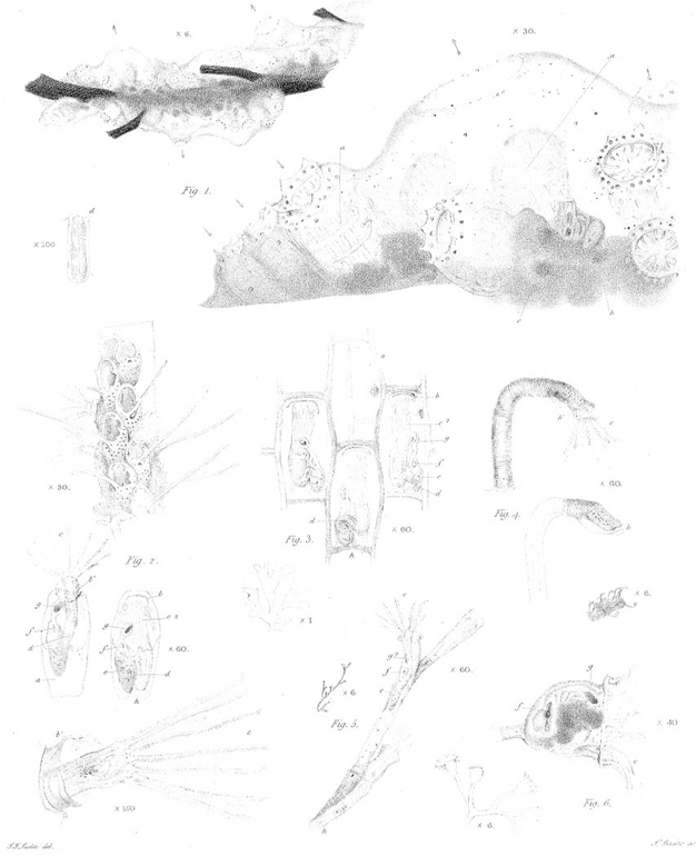
Fig. 1. Tubularia indivisa (page 366.). The arrows show the direction of the currents within.
c. The place of their return in the tube.
d, e. Nodous parts.
a. The mouth of the polypus, pressing its fluid contents into the stomach b.
a 2. The same on the flow returning from the stomach.
a 3. The mouth swollen, and the stomach emptied.
f. Original end of the shell.
a 4. The tube and shell extending to the hinder arms, which are in the process of destruction.
a 5, a 6. Appearances at the end of the tube at two later periods.
Fig. 2. Sertularia pluma (page 369.).
X 1. Anastornosis of the creeping tube and manner of growth.
X 6. A plume.
a. Side of the cells.
b. Back of the same.
Fig. 3. Sertularia pumila (page 371.).
a. Septum in the cells.
a 2. The same, seen from above by section of the shell.
b. The aperture in the septum, by which the polypus is connected with the tube.
c. Mouth of the polypus.
d. Stomach.
e. Empty ovary.
Fig. 4. Sertularia setacea (page 371.). The soft matter occupies one side of the stem, with a current of particles flowing in its axis.
a. Septum in the small cell.
b. Spiny ovaries, with the ova indistinctly seen.
Fig. 5. The minute Sertularia described page 372, with the appendage on the side of its cells.
a. Its large ovary, containing an opake mass, connected with the base and end by a column, and with the sides by strings b.
PLATE IX.
A young stem of Campanularia dichotoma? with its annular strictures (page 373.).
a. A branch terminated by a perfect polypus.
[page break]

[page break]

[page break]
[page] 387
b. Another, of which the polypus, with a deformed shell, is recently completed. X 200. The ends of two of its arms, more enlarged.
c. A sprout in the act of growth.
c 2. The same sprout six hours later. The arrows show the course of the currents within.
c 3. The same after six hours more: rudiments of a shell forming.
c 4. Further growth of the same in eight hours. The diverging lines at the end of c, c 2, c 3, &c. accompany the forming process.
c 5 to 9. Progress of the imperfect cell and polypus towards completion.
a 2 to 5. Stages in the absorption of the polypus a.
PLATE X.
Fig. 1. Campanularia dichotoma? with ovaries in different states (page 375.).
a. An immature one, of which the shell appears to be in progress of growth at the end.
b. One more advanced.
c. Another, which has begun to protrude its ova.
b 2, b 3. Exclusion of the first ovum from the ovary b.
b 4. The young polypus discharging its active particles.
b5. Its appearance after their discharge, before the emergence of the second ovum.
b 6. The same ovary after the first and second young polypus had been absorbed, when the third was become rugose, and the fourth had just discharged its particles.
c 2. The ovary c with the columella remaining, before discharging its last ovum.
Fig. 2. The simple Campanularia described page 376.
Fig. 3. Coryne (page 376.).
a. Its small cell, in the axis of which the alternate current was seen.
b. The naked ova, in the peduncles of which the same current appeared.
PLATE XI.
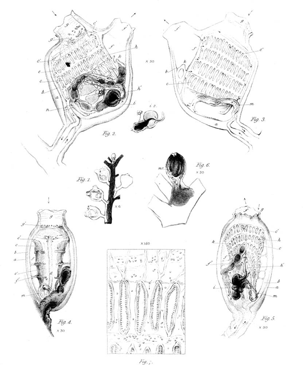
Animals of the Ascidia described page 378. The interior is seen through the transparent coats.
Fig. 1. Manner of their growth.
Fig. 2. The right side of one supported on its peduncle.
Fig. 3. The left side of another.
Fig. 4. The back.
Fig. 5. The front and base.
Fig. 6. A sprout ending in the rudiment of an animal.
The letters refer to the same parts in all. The small arrows denote the course of the circulation when the forward impulse is given by the heart; the large arrows at the orifices, the influx and efflux of water.
a. The envelope.
b. The mantle.
[page] 388
c. The branchial sac, with its four rows of spiracles.
c′ c′ c′. Its line of junction with the mantle above and behind.
d. Vertical dorsal stripe. (fig. 4.)
e, e. Finger-like processes projecting from the branchial sac into the cavity of the mouth within. (fig. 2, 4.)
f. Cavity between the sac and mantle, ending in the funnel f′.
f″. Ligaments stretched between the sac and mantle. (fig. 4, 5.)
g. The oral opening.
g′. Its tentacula. (fig. 2, 4.)
h. The downward stream of food which flows into the œsophagus h′. (fig. 2, 5.)
i&i 2. The stomach, its fore part enveloped by the liver? (fig.2, 5.)
k. The vent terminating the intestinal canal.
l. Lacteals? uniting near a mass of transparent globular bodies, with which two lobes are connected. (fig. 2.) The latter are wanting in some individuals.
m. The heart.
n. Point where the back stream of blood communicates with the heart when the peduncle is severed. (fig. 2.)
Fig. 7. A portion of the branchial sac, more magnified, to show the ciliæ surrounding the spiracles and the particles of the blood.
PLATE XII.
Fig. 1. Polyclinum (page 382.), with a portion of the same more magnified. The branchial sacs, &c. are seen through the transparent common coat. The arrows pointing inwards indicate the oral openings; those pointing outwards, the common funnels.
a, a. Ledges on the interior of the branchial sac, each ending at a moveable spiral process in front, seen in two of the animals.
b. The heart of one of them.
c. Vent of the same, with other viscera imperfectly seen.
d. Appearance of a spiracle when the ciliæ are closed.
Fig. 2. Flustra pilosa, encrusting a fucus, and single animals of the same, seen through the shell and coat (page 384.).
a. The shell.
b. Valve in the coat, through which the ciliated arms c, and the mouth are protruded.
b′. The short sheath.
c 2. The arms when drawn in, with the neck d folded back.
e. Pouch (containing the stomach, liver, &c.?).
f. Place of gyration of particles in the intestine.
g. Rectum.
h. Ligaments or muscles between the animal and the base of the cell.
Fig. 3. Flustra papyracea (page 384.). The animals folded in their cells. The letters in this and the following figures refer to the same parts as in the last.
Fig. 4. Anguinaria anguina (page 385.).
Fig. 5. Tibiana (page 385.).
Fig. 6. A zoophyte described page 385.
[page break]