[page] 239
The Action of Carbonate of Ammonia on the Roots of certain Plants. By CHARLES DARWIN, LL.D., F.R.S.1
[Read March 16, 1882.]
MANY years ago I observed the fact that when the roots of Euphorbia Peplus were placed in a solution of carbonate of ammonia a cloud of fine granules was deposited in less than a minute, and was seen travelling from the tip up the root from cell to cell*. The subject seemed to me worthy of further investigation. Plants of the same Euphorbia were therefore dug up together with a ball of earth, and having been left for a short time in water, the roots were washed clean. Some of the finer transparent rootlets were then examined, and sections were made of the thicker roots, generally by my son Francis, who has aided me in many ways. All the cells were found to be colourless and destitute of any solid matter, the laticiferous ducts being here excluded from consideration. These roots, after being left for a few minutes or for several hours in solutions of different strengths, viz. from 1 to 7 parts of the carbonate to 1000 of water, presented a wonderfully changed appearance. A solution of only 1 part to 10,000 of water sufficed in the course of 24 hours to produce the same result. In well-developed cases the longitudinal rows of cells close to the tip of the root, with the exception of those forming the extreme apex, were filled with brown granular matter, and were thus rendered opaque. Long-continued immersion in water produced no such effect. The granular masses were square in outline, like the cells in which they were contained; but they often became rounded after a day or two; and this was apparently due to the contraction of the protoplasmic utricle. Above the dark-brown cells, which form a transverse zone close to the tip, and which apparently corresponds with the zone of quickest growth, the roots, as seen under a high power, are longitudinally striped with darker and lighter brown. The darker tint is due to the presence of innumerable rounded granules of brownish matter; and the cells containing them are arranged in longitudinal rows, while other longitudinal rows are destitute of granules. In a few instances the rows differed slightly in tint, and yet no
* 'Insectivorous Plants,' 1875, p. 64. The subject was at that time, 22 years ago, only casually investigated; and I believe that I erred greatly about Lemna, unless, indeed, some different species was then observed, or that the season of the year makes a great difference in the behaviour of the roots, which is not probable.
LINN. JOURN.—BOTANY, VOL. XIX. X
1 Read by Francis Darwin. An abstract of this article and Darwin 1882, F1801, was published by Francis Darwin: The action of ammonia on the roots of certain plants and on chlorophyll bodies. Nature 25 (23 March 1882): 489-90. A2972
[page] 240
granules could be seen in the darker cells; and I suppose that this was owing to their being too minute to be visible. Occasionally, in the upper parts of the roots, the granules became confluent, and formed one or two small rounded masses of hyaline brown matter. The striped appearance sometimes extended from the tips of the finest rootlets close up to the stem of the plant.
On a casual inspection it would be said that the longitudinal rows of brownish and of almost colourless exterior cells regularly alternated with one another; but on closer examination, two or three adjoining rows of cells were often seen to contain granules, and in other places two or three ordinary rows contained only colourless fluid. In one instance many adjoining longitudinal rows contained granules; but the tendency to alternation was even here well shown, as the alternate rows differed in tint from including a greater or less number of granules. High up the roots the alternations often quite failed, as all the exterior cells contained granules. If a longitudinal row of cells with granules is traced up a rootlet, it is seen to be soon interrupted by one or more colourless cells; but I have traced as many as 18 cells in a row all containing granules. So, again, a longitudinal row of colourless cells changes after a time into one with granular matter. As a root thickens upwards, some of the longitudinal rows of cells divide into two rows; and a row containing granules may divide into two such rows, or into one with and another without granules; and so it is with dividing rows of colourless cells. I could not perceive the least difference in shape or size, or in any other character, between the cells of the same rank which contained and those which were destitute of granules.
Near the tip of the root it is the exterior cells which become charged, after immersion in the solution, with brown granular matter; and this often holds good with the cells of the root-cap. Higher up the root, the layer of cells formed by the alternating longitudinal rows with and without granules is sometimes bounded externally by a layer of empty cells, which, I suppose, had by some means been emptied of their contents, and were ready to be exfoliated. Besides the exterior cells with and without granules, many separate cells in the parenchyma at different depths from the surface, and all or several of the elongated endoderm-cells surrounding the central vascular bundle, are more or less filled with granular matter, none of which cells contained any solid matter before the roots were immersed in the solution.
[page] 241
I should have felt little surprise at the effect produced by the solution if all the cells of the same nature (for instance, if all the exterior cells or all the parenchyma-cells) had been equally affected. The strong tendency to alternation in the exterior cells is more especially remarkable. There is also another remarkable fact with respect to these latter cells, namely, that those containing the granules do not give rise to root-hairs, as these arise exclusively from the colourless and apparently empty cells. In longitudinal sections of one root, 62 hairs were traced down to such colourless cells; and I was not able to find a single one arising from a cell which contained granules. But I shall have hereafter to return to this subject.
With respect to the rate at which the granular matter is deposited, if a rootlet is placed under a cover-glass and irrigated with a few drops of the solution, some deposition occurs before the slide can be transferred to the microscope and the focus adjusted. A thin rootlet was therefore arranged for observation, and a drop of the solution (7 to 1000) placed on the edge of the cover-glass, and in 20 seconds the cells near the tip became slightly clouded. Another thin rootlet was placed with the tip projecting beyond the cover-glass, and the focus was adjusted to a point at a distance of .07 inch from the tip, on which a drop of the solution was then placed, and the cells at the above distance became cloudy in 2 m. 30 sec.
Various other solutions, beside that of carbonate of ammonia, caused the deposition of granules in the same cells as in the foregoing cases. This occurred conspicuously with a solution of 4 parts of phosphate of ammonia to 1000 water; but the action was not so rapid as with the carbonate. The same remarks are applicable to nitrate of ammonia. A solution of one part of fuchsine, which contains nitrogen, to 50,000 of water distinctly acted. A solution of 2.5 parts of pure carbonate of soda to 1000 water caused, after 24 hours, the cells close to the tip to become very brown from being charged with fine granular matter; and higher up the rootlets, longitudinal rows of cells, either containing coarse granules or pale-brown fluid without any distinguishable granules, alternated with rows of colourless cells. Lastly, roots immersed for only one hour in a watch-glass of water, to which two drops of a 1-per-cent. solution of osmic acid had been added, presented an extraordinary appearance; for the exterior cells in alternate rows, some parenchyma, and most of the endoderm-cells contained much almost black granular matter.
X 2
[page] 242
The granules precipitated through the action of carbonate of ammonia are never afterwards, as far as I could judge, redissolved. Roots still attached to living plants were immersed in solutions of 1 part of the carbonate to 500, to 2000, and to 4000 parts of water, and granular matter was deposited in the cells in the usual manner. The roots were then left in damp peat or in water, with the stems and leaves exposed to the air and light, for various periods between 2 and 15 days. The roots were then reexamined at different times, and granules were found in almost every instance in the cells. But it should be noticed that though the plants themselves looked healthy, the finer roots were flaccid, and sometimes showed evident signs of decay; so that it was manifest that they had been much injured by the treatment to which they had been subjected, probably by their immersion in the solution.
With respect to the nature of the granules, I can say but little. They were not dissolved by long-continued immersion in alcohol or in acetic acid, or by irrigation with sulphuric ether. They were not dissolved by a 10–per-cent. solution of common salt, which was tried at the suggestion of Mr. Vines,1 who has found that this solution dissolves aleurone-grains either partially or completely. When sections or rootlets containing freshly deposited granules were left for a day or two in glycerine and water, these were sometimes broken up, so as to be no longer visible, and the cell-sap in this case acquired a brownish tint. When sections or thin rootlets were heated for a short time in a moderately strong solution of caustic potash, and afterwards left in it for a day or two, the granules were dissolved; whereas the hyaline globules in the laticiferous ducts were not dissolved. From these several facts I suppose that the granules are of the nature of protein.
After roots had been left for 2 or 3 minutes in water heated to a temperature of 210°–212° F., and were then placed in a strong solution of the carbonate of ammonia, no granular matter was deposited; and this seems to indicate that the action is a vital one. On the other hand, granules were often deposited in the cells, even the loose cells, of the root-cap, and it is very doubtful whether these could be alive. I may add that these root-cap cells were coloured, by a weak solution of fuchsine, of a brighter pink than those in other parts of the rootlets.
Other Euphorbiaceous Plants.—The exterior cells of the roots of Euphorbia amygdaloides were much less acted on (Nov. 16) by
1 Sydney Howard Vines (1849-1934), botanist and Fellow of Christ's College, Cambridge, 1876.
[page] 243
a solution of carbonate of ammonia than those of E. Peplus. Here and there two and three cells in a row contained brownish granules, and these abounded in the elongated endoderm-cells. Nearly the same remarks are applicable to E. myrsinites, though in most specimens the cells with granules were still rarer. The roots of two fleshy species, E. rhipsaloides and ornithopus, did not appear to be at all affected by the solution.
Turning now to other Euphorbiaceous genera, the roots of Poinsettia pulcherrima, Manihot Glaziovi, Croton oblongifolium, and Hevea Spruciana were not affected. Nor were those of Mercurialis perennis, as far as the exterior cells are concerned; but here and there a single cell in the parenchyma became blue; but these cells were not carefully examined*. Judging from the cases presently to be given, they probably contained granules which had been precipitated by the ammonia solution.
On the other hand, the roots of Phyllanthus compressus were conspicuously acted on by an immersion of 21 hours in a solution of 4 parts of the carbonate to 1000 of water, though in a somewhat different manner from those of Euphorbia Peplus. In parts the exterior cells in many adjoining longitudinal rows contained brownish granules; while in other parts at no great distance many adjoining rows were colourless and empty—that is, contained no solid matter. For instance, in one place 13 longitudinal rows with granules ran alongside one another, then came a single row of empty cells, and then at least 9 rows with granules. In another place there were 13 adjoining rows of cells all empty. When one of these rows was followed up or down the root for some distance, it changed its character, either becoming or ceasing to be granular, and then resuming its former character. Close to the tips of the roots all the longitu-
* The rhizomes and buried parts of the stems of this plant are white; but after immersion for a day in the ammonia solution they became in parts either pale or rich blue. This change of colour occasionally occurred in parts exposed to the air which had not been subjected to the solution. As a similar change occurs in certain cells in the roots of various plants after their immersion in the solution, I asked Mr. Sorby1 to be so kind as to examine the rhizomes and underground stems of the Mercurialis. He informs me that he does not understand the change of colour; but he was unable to spare time for a full examination. He found that when the rhizomes and stems were boiled in alcohol, they yielded matter which was soluble in water, and which appeared to pass so rapidly into a brown substance with curious shades of green, that the real change was hidden. On the whole, the appearances differed a good deal from those observed by him in the case of blue flowers.
1 Henry Clifton Sorby (1826-1908), geologist who pioneered microscopic petrology. Sorby was part of the deputation of the Yorkshire Naturalists' Union which presented a memorial to Darwin in 1880. See Darwin 1880.
[page] 244
dinal rows of cells contained brownish matter; but this matter in several instances consisted of small dark-brown spheres, due apparently to the aggregation of granules. The endoderm-cells round the vascular bundle contained either similar spheres or granular matter.
As many adjoining rows of cells on the surface of the roots of this plant had the same character, an excellent opportunity was afforded for observing the relation of the root-hairs to the cells; and in several dissected roots it was manifest that, as a general rule, the hairs rose exclusively from the colourless empty cells; whereas none arose from those containing granules. Twice, however, partial exceptions to this rule were observed: in one case the exterior walls of two adjoining cells, and in another case those of four adjoining cells, projected, so that they formed short blunt papillæ which included granules; and these papillæ exactly resembled nascent root-hairs. It is not, however, certain that they would ever have become fully developed.
All the exterior cells close to the tip of the root in this case and in many others contained matter which was acted on by carbonate of ammonia; and I was led by various appearances to suppose at one time that this matter remained in all the higher cells until it was consumed in some of them by the formation of the root-hairs. These consequently would arise exclusively from cells in which no granules would be deposited when they were acted on by the solution. In opposition to this supposition is the fact, first, that root-hairs could be seen beginning to be developed from empty cells; and, secondly, that very many cells which were empty apparently had never produced root-hairs. Nor does this notion throw the least light on single cells in the parenchyma and on many cells, though not all, in the endoderm containing granular matter.
With another Euphorbiaceous plant, Cœlebogyne ilicifolia, the immersion for 20 hours of its roots, or of thin sections of the roots, in a solution of 4 parts of carbonate of ammonia to 1000 parts of water produced a singular effect; for many separate cells in the parenchyma and those in the endoderm surrounding the vascular bundle assumed a pale or dark blue, and sometimes a greenish colour. As far as I could judge, both the granules within these cells and the cell-sap became thus coloured. Irrigation with sulphuric ether did not affect the colour, though the many oil-globules in the cells were dissolved.
[page] 245
The foregoing observations on the Euphorbiaceæ led me to experiment on the roots of some other plants belonging to various families. At one time I erroneously imagined that there was some relation between the deposition of granules in certain cells and the presence of laticiferous ducts, and consequently an undue number of plants with milky juice were selected for observation. A solution of carbonate of ammonia produced no obvious effect on the roots of a small majority of the plants which were tried; but on several a slight, and on others a marked, effect was produced. I should state that when the exterior appearance of a root did not indicate any action, sections were rarely made; so that the interior cells were not examined. No obvious effect was produced with the following plants:—Argemone grandiflora, Brassica oleracea, Vicia sativa, Trifolium repens, Vinca rosea, Hoya campanulata, Stapelia hamata, Schubertia graveolens, Carica Papaya, Opuntia boliviensis, Cucurbita ovifera, a Begonia, Beta vulgaris, Taxus baccata, Cycas pectinata, Phalaris canariensis, a common pasture-grass, Lemna, and two species of Allium. It may perhaps be worth notice that the radicles, but not the hypocotyls, of seedlings of Beta vulgaris were completely killed by an immersion for 20 hours in solutions of either 4 or of only 2 parts of the carbonate to 1000 of water; and this occurred with no other plant which was tried.
With the following plants the solution produced some slight effect. The roots of a fern, Nephrodium molle, were immersed for 20 hours in a solution of 4 to 1000; and this caused the deposition of some brown granular matter in the cells near their tips; and more or less confluent globules could be seen in the underlying parenchyma-cells. So it was with an unnamed greenhouse species of fern; and in this case the almost loose cells of the root-cap contained brown granules. The roots of a Ranunculus (R. acris ?) similarly treated exhibited near their tips brown granular matter. The tips also of the roots of Dipsacus sylvestris became, under similar treatment, almost black; and higher up the roots, here and there a single parenchyma-cell was coloured pale blue. This occurred in one instance when a rootlet was looked at 35 minutes after irrigation with the solution. Several roots of Apium graveolens were left for 20 and 24 hours in solutions of 4 and 7 to 1000; and in some cases brownish granules, more or less aggregated together, were deposited in some of the exterior cells, and a few of the deeper cells in the parenchyma
[page] 246
were coloured blue. The tips of the roots of Pastinaca sativa turned dark brown by a similar immersion; but this was due to the formation of orange-brown balls of matter near the vascular bundle; higher up the roots there were no granules in the exterior cells. The tips of the roots of Lamium purpureum, after an immersion of 18 hours in a solution of 4 to 1000, were rendered brown, and the cells contained innumerable pale-coloured hyaline globules. The older roots of Leontodon Taraxacum and of a Sonchus had their tips turned brown by the solution. With Lactuca sativa the tips were rendered opaque; but much granular matter was not deposited except in that of one rather thick leading root, and here short longitudinal rows of cells containing dark-brown granular matter alternated with rows of colourless cells; the almost loose cells of the root-cap likewise contained brown granules. In the several following cases a much more strongly marked effect was produced by the solution.
Urtica.—This plant, the common nettle, shall be first considered, as it is distantly allied to the Euphorbiaceæ, though the roots are not so much affected as in succeeding cases. Several roots were left for 27 hours in a solution of the carbonate (4 to 1000). In one of them the exterior cells were plainly tinted of a brown colour in many longitudinal rows, but they contained no visible granules; and these rows regularly alternated with others formed of colourless cells. In another part of this same root all the exterior cells were coloured dark brown, and contained visible granules, which were generally collected into heaps at one end of the cell, or were fused together in some instances into small brown spheres. In a second, rather thick root, there was a space in which all the exterior cells had become brown; but at no great distance rows of brown and colourless cells regularly alternated. In a third, rather thick, and in a fourth, thin root the alternation was extremely regular. Near the tip of a fifth (thin) rootlet two rows of a brown colour ran alongside one another in many places; but when these and other single rows were traced up the root, they changed into colourless rows, and afterwards reassumed their former character. Whenever the root-hairs were traced down to their bases, they were seen to arise from colourless cells. Neither granules nor brown fluid were observed in the parenchyma-cells nor in those surrounding the vascular bundle.
Some roots which had been left in water for several days were
[page] 247
longitudinally striped with very faint brown lines; and one cell was observed which included granules; so that plain water produces some effect. These same roots, after being irrigated with a solution of 7 to 1000, were left for 24 hours; and now the longitudinal rows of brown cells had become much darker, and presented a much stronger contrast with the colourless cells. Several of the brown cells moreover included granules, which here and there were aggregated into small dark-brown rounded masses.
Drosera, Dionæa, and Drosophyllum.—The roots of the plants belonging to these three closely allied genera are strongly acted on by a solution of carbonate of ammonia. In the case of a young plant of the Dionæa, all the exterior cells of the roots, after immersion for 24 hours in a solution of 4 to 1000, contained almost black or orange, or nearly colourless spheres and rounded masses of translucent matter, which were not present in the fresh roots. In this case, therefore, the exterior cells did not differ in alternate rows. Near the extremity of one of these roots many separate cells in the parenchyma, as seen in transverse sections, contained similar translucent spheres, but generally of an orange colour or colourless. The cells surrounding the vascular bundle abounded with much smaller dark-coloured spheres.
Three main or leading roots of Drosophyllum lusitanicum were cut off and examined before being immersed in the solution, and no aggregated masses could be seen in them. Two were left for 22 hours in a solution of 4 to 1000, and they presented an extraordinarily changed appearance; for the exterior cells in many rows from the tips to the cut-off ends of the roots included either one large, or, more commonly, several spherical or oval, or columnar masses of brown translucent matter. The columnar masses had sinuous outlines, and appeared to have been formed by the confluence of several small spheres. The loose, or almost loose, oval cells composing the root-cap included similar brown spheres; and this fact deserves attention. Two rows of cells containing the just-described masses often ran up the root alongside one another; and sometimes there were three or four such adjacent rows. These alternated with others which were colourless, and contained either no solid matter, or rarely a few minute pale spheres. These roots were carefully examined; and all the many root-hairs arose from the colourless rows of cells, except in some few cases in which the cells on both sides abounded to an unusual
[page] 248
degree with aggregated masses; and here root-hairs arose from cells including a very few minute spheres.
In longitudinal sections of the above roots, the cells in the parenchyma at different depths from the surface were seen to include spheres, but many of them were of small size and pale-coloured. There was no marked increase in the amount of aggregated matter in the cells closely surrounding the vascular bundle, as is so often the case with other plants.
The third cut-off root was placed under the microscope, and was irrigated with a solution of 7 to 1000. After 13 minutes very small translucent granules could be seen in many of the cells; and after 35 minutes several cells near the cut-off end contained moderately large spheres of translucent matter. But I suppose that the solution was too strong; for the granules disappeared after about 45 minutes, except close to the tip; and the higher parts of the root no longer presented a striped appearance. Nevertheless the large spherical, oval, and oddly shaped masses in the cells near the cut-off end remained perfect, and they were watched for the next 2¼ hours. During this time they slowly changed their shapes, but not afterwards, though observed for nearly 24 hours. For instance, two spheres in one cell became confluent and formed an oval mass; two other spheres ran together and formed a dumbbell-shaped body, which ultimately changed into a sphere; and, lastly, an irregular mass first became oval, then united itself with another oval mass, and both together became spherical.
Saxifraga umbrosa.—This plant, from its affinity to the Droseraceæ, was cursorily observed. Many of the exterior cells of roots which had been immersed for 19 hours in a solution of 4 to 1000 were filled with brown granular matter. Only two or three cells in a longitudinal row were thus filled; but sometimes four or five such short rows were grouped together; and these groups alternated with rows of colourless cells.
Sarracenia purpurea.—Two rootlets were left in water for 24 hours, but they presented no granules or aggregated masses. They were then irrigated with a solution of 7 to 1000, and in 20 minutes pale-brown aggregated masses could be distinctly seen near their tips. Two other, almost colourless, rootlets were left for 1 hour 10 minutes in the same solution; and now all the exterior cells contained brown granular matter, but much darker in some cells than in others. Some of the cells contained,
[page] 249
besides the granules, oval and occasionally spherical masses of transparent, almost colourless matter, which apparently did not change their shapes. The cells round the central vascular bundle included similarly shaped masses, but of a yellowish-brown colour. These roots and others were left for 24 hours in the solution of 7 to 1000, and their tips were now blackened. Some of the exterior cells, more especially those of the thicker roots, were filled with orange instead of brown granules; while other cells contained oval, spherical, or oddly shaped masses of orange, instead of almost colourless or pale-brown translucent matter. Some of these masses consisted of an aggregation of small, partially confluent spheres of different tints of orange. In transverse sections it could be seen that the two exterior layers of cells and those surrounding the vascular bundle contained the above-described masses, while the more central parenchyma-cells abounded with grains of starch. A solution of 4 parts of the carbonate to 1000 of water sufficed to produce similar effects.
The root-hairs, after immersion in the solution, were not so transparent as is commonly the case, from including very fine granular matter, and from their shrunken protoplasmic lining being of a yellowish colour. The roots themselves were also usually opaque. Consequently the root-hairs were not easily traced down to their bases. They were distributed very unequally, being quite absent from the browner parts of the roots, while present on the parts which had remained pale-coloured. Notwithstanding this latter fact, it is very doubtful whether the rule of root-hairs arising almost exclusively from cells destitute of solid matter here holds good.
Pelargonium zonale.—A fresh root was examined, and the cells contained no granules. It was then irrigated with a solution of 7 to 1000, and in about 15 minutes granules could be distinctly seen in the exterior cells in alternate rows. Two other rootlets, after being left in water for 48 hours, were not at all affected. They were then irrigated with the same solution and reexamined after 24 hours; and now the exterior cells in rows, as well as those surrounding the vascular bundle, abounded with granular matter. Other roots were left for 48 hours in a solution of 4 to 1000; and the cells near their tips were so packed with dark-brown granular matter as to be blackened. Higher up the roots, the granules were pale brown, translucent, irregularly rounded, and often more or less confluent. In some dark-coloured rootlets the
[page] 250
cells included a few small spheres of dark brown matter instead of granules. Usually the cells containing the granules formed single longitudinal rows, which alternated with rows of colourless cells. But occasionally several adjoining rows included granules; thus in one place two adjacent rows of cells with granules were succeeded by an empty row; this by two alternations of granules and empty rows; then came two adjoining rows with granules, an empty row, and three adjoining granular rows. In another place an empty row was succeeded by five adjoining rows with granules; these by an empty row; this by three adjoining rows with granules, and this by an empty row.
After many casual observations, in which all the root-hairs appeared to arise from cells destitute of granules, this was found to be the case with 50 hairs which were traced down to their bases. With one problematical exception, not a single hair could be found which arose from a cell containing granules. In this one exceptional case, a hair seemed to spring from the transverse wall separating two cells; but with a good light and under a high power, the wall apparently consisted of two walls, separated by an excessively narrow clear space, as if a cell had here failed to be fully developed.
The solution likewise caused the precipitation of granules in the elongated cells surrounding the vascular bundle, and in some tubes or ducts within the bundle. The solution apparently does not act on cells which have been killed. The ends of a root were torn open, so that the vascular bundle was fully exposed; the root was then left for 24 hours in a strong solution of 7 to 1000, and no granules were deposited in the exposed cells round the vascular bundle; but by tearing open fresh parts of the same roots, these cells were found full of granules.
The granules were not dissolved by immersion for 24 hours in alcohol; but they were dissolved by a cold solution of caustic potash. The dissolution, however, took place very slowly; for though on two occasions the granules wholly, or almost wholly, disappeared after an immersion of 20 hours, yet with a thicker root they were not dissolved, though rendered browner, by an immersion for this length of time; but they finally disappeared after 18 additional hours in a fresh solution of the potash. In the cells round the vascular bundle, from which the granules had been dissolved by the potash, matter resembling oil-globules in appearance remained.
[page] 251
Lastly, two drops of a 1-per-cent. solution of osmic acid were added to ½ oz. of distilled water, and some roots were left in this fluid for 20 hours. They were affected in very different degrees. Some were only a little discoloured; and in such roots a single exterior cell here and there contained either blackish granules or small black spheres. Other roots were much blackened; and in these longitudinal rows of dark brown or blackened cells plainly alternated with colourless rows. The cells surrounding the vascular bundle and many of the parenchyma-cells also contained blackened granules. Hence it is probable that carbonate of ammonia likewise acts on some of the parenchyma-cells; but if so, the fact was overlooked, or accidentally not recorded, in my notes.
Oxalis Acetosella.—Roots were first examined, and then placed in a solution of 7 to 1000. Some slight degree of aggregation was seen in a few minutes. After 30 minutes all the cells near the tips contained rounded accumulations of granules. Higher up in one of the roots, single cells, or from two to five cells in a row, were filled with minute hyaline globules. In some places these had become confluent, so that they formed larger globules having a sinuous outline. The cells underlying the exterior layer likewise contained extremely fine granular matter. Still higher up the same root there were considerable spaces in which none of the cells contained granules. But again higher up, the granules reappeared. The root-hairs were numerous; but not one was seen which arose from a cell containing granules.
Roots of Oxalis sepium, comiculata, and of a greenhouse species with small yellow flowers were immersed in a solution of 7 to 1000, and granular matter was deposited in the layer of cells underlying the exterior layer. This occurred in the case of O. sepium in 20 minutes. With O. corniculata the cells with granules were isolated—that is, did not form rows; and the granules were either brown or of a bluish-green colour. In the case of Oxalis (Biophytum) sensitiva, the exterior cells of the roots, after immersion for 44 hours in the same strong solution, were not much affected; but some of the deeper parenchyma-cells contained dark-brown translucent spheres, and the elongated cells round the vascular bundle were almost filled with granular matter.
Fragaria (garden var. of the common strawberry).—Some white, almost transparent roots from a runner were examined (Dec. 12), and the cells contained no solid matter, except starch-
[page] 252
grains. They were then irrigated with a solution of 7 to 1000; and in from 10 to 15 minutes they became very opaque, especially near their tips. After being left a little time longer in the solution, longitudinal sections were made. The cells forming the exterior layer contained no solid matter, but the walls had become brown. There was much brown, finely granular matter in many of the parenchyma-cells at different depths from the surface; and these formed interrupted longitudinal rows, which alternated in the same zone with rows of empty colourless cells. Almost all the endoderm-cells likewise contained granules. In the parenchyma the cells which included much granular matter contained no starch-grains; while those abounding with starch-grains contained only a few or no granules. The fact was best seen after the sections had been irrigated with a solution of iodine; and they then presented a very remarkable appearance, considering how homogeneous they had been before being treated with the ammonia and iodine; for the fine granular matter was rendered still browner and the starch-grains of a beautiful blue. These roots were left for a week in diluted alcohol, and the granules were not dissolved.
Not a single root-hair could be found on these roots. A rooted stolon was therefore dug up and potted on Dec. 12th; it was then forced forwards in the hot-house, and afterwards kept very dry. When examined on Jan. 3rd the roots were found clothed with innumerable root-hairs; and they were then left for 23 hours in the solution of 7 to 1000. Sections of the thicker roots presented exactly the same appearances as described above; and the exterior cells, from which the root-hairs arose, were destitute of granules. The thinner roots differed somewhat in appearance, as the parenchyma-cells did not contain any fine granules, but in their places small, spherical, or oval, or irregularly-shaped masses or filaments of brown translucent matter, resembling a highly viscid fluid. There were also in these cells other still smaller colourless spheres. The cells, however, close to the tip of the root were filled with brown granular matter.
Solanum (capsicastrum ?, var. Empress).—Roots, after an immersion of 20½ hours in a solution of 4 to 1000, were split longitudinally and examined, but with no great care. The exterior cells did not appear to have been affected; but some of the parenchyma-cells close beneath the exterior cells contained minute aggregated masses of brown, opaque, or sometimes hyaline granules. More-
[page] 253
over many, but by no means all, of the elongated cells surrounding the vascular bundle included dark-brown fine granular matter. Three roots which had been left in water for the same length of time, viz. 20½ hours, were similarly examined, but their cells presented none of the above appearances.
Primula acaulis.—Several roots were left (Dec. 22) for 18 hours in a solution of 4 to 1000, and they were all much affected, except some of the thinnest rootlets. Many of the exterior cells contained granules within the shrunken protoplasmic utricle, which had contracted into one, two, or even three, oval or spherical bags, lying within the same cell. The rows of cells containing the granules showed some tendency to alternate with rows of empty cells. The granules were rendered orange-brown by iodine. The innumerable root-hairs all arose from the empty cells; and I saw only two partial exceptions, in which the outer walls of cells containing granules were produced into short papillæ, as in the formerly described case of Phyllanthus, and these resembled nascent root-hairs. Within one of these papillæ, granules surrounded by the shrunken utricle could be seen. In the parenchyma single cells were seen containing minute hyaline globules, which were colourless or pale or dark blue, or occasionally greenish or yellowish. Many of the endoderm-cells likewise contained more or less confluent hyaline globules; but these were colourless, and larger than those in the parenchyma. They resembled starch-grains so closely that they were tried with iodine, but were not coloured blue. Roots which had been kept for 48 hours in water exhibited none of the coloured or colourless globules; but these appeared when the roots were afterwards immersed for 24 hours in the ammonia solution.
Although it is certain that granules were deposited in the exterior cells in the case just described, yet in four other roots, after an immersion of 24 hours in the solution, no granules could be seen in any of the exterior cells. Some of the parenchyma-cells, however, were of a fine blue colour, and contained many globules or granules, but no starch-grains, while others contained starch-grains as well as some few globules.
Cyclamen persicum.—Sections taken from roots of this plant which had been immersed in a solution of carbonate of ammonia presented an extraordinary different appearance from those of fresh roots. All the cells in the latter appeared empty, excepting those of the endoderm, which sometimes included a few very fine pale-
[page] 254
Fig. 1.
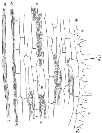
Longitudinal section of root of Cyclamen persicum after immersion in a solution of carbonate of ammonia, and deposition in some of the cells of granules. a, part of vascular bundle; b, endoderm-cells; c, parenchyma-cells; d, exterior cells of the root, bearing root-hairs; e, with their tips cut off. Drawing made by aid of a camera, magnified 260 times, but here reduced to two thirds of original scale.
Fig. 2.
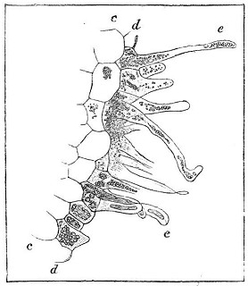
Transverse section of another part of the same root, magnified as before, showing the exterior cells, d, together with the root-hairs, e, here containing granules.
1 See the pencil drawings by Francis Darwin these woodcuts are based on in CUL-DAR28.2.A1a-b Images and Darwin to J. D. Cooper 4 February 1882 and 22 February 1882. Correspondence vol. 30, pp. 58, 93.
[page] 255
coloured granules, unlike those in the same cells after immersion. Thick and thin roots were left for 22 hours in a solution of 7 to 1000, and the cells forming the exterior layer were filled over considerable spaces with green granules, while over other spaces they were empty. The granular and empty cells did not form regular alternate rows, as occurs in so many other plants; yet, as we shall presently see, there is occasionally some degree of alternation. The exterior cells with the green granules were so numerous in certain cases, that roots which had been pale brown before immersion were afterwards distinctly green. The green granules sometimes became aggregated into spherical, or oval, or elongated masses having a sinuous outline; and some of these are shown within the root-hairs in fig. 2. Many of the cells of the parenchyma, either standing separately or two or three in a row (as shown in fig. 1), contain similar green, or sometimes brownish, granules. Almost all the narrow elongated cells of the endoderm (b, fig. 1) likewise contain these granules, with merely here and there an empty cell. Although both kinds of cells often appear as if gorged with the granules, yet these really form only a layer adhering to the inside of the protoplasmic utricle, as could be seen when cells had been cut through. With some thick fleshy roots, after an immersion for 42 hours (and thick roots require a long immersion for the full effect to be produced) the green granules in the parenchyma-cells had become completely confluent, and now formed spheres of transparent green matter of considerable size.
The granules are not dissolved, nor is their colour discharged by sulphuric ether. Acetic acid instantly changes the green into a dull orange tint. The granules are not dissolved by alcohol. Their precipitation by the ammonia solution seems to depend on the life of the cell; for some transverse sections were examined and found colourless, as well as destitute of granules. They were then irrigated with a solution of 7 to 1000, and reexamined after 22 hours; and only a very few cells in two out of the five sections showed any trace of colour, which, oddly enough, was blue instead of green. The few coloured cells occurred exclusively in the thickest parts of the sections, where the central ones would obviously have had the best chance of keeping alive for some time. In these coloured cells a little very fine granular matter could be distinguished.
On most of the roots root-hairs were extremely numerous,
LINN. JOURN.—BOTANY, VOL. XIX. Y
[page] 256
and they generally arose from cells destitute of granules; yet in many places whole groups of cells abounding with granules gave rise to well-developed root-hairs. Therefore the rule which holds good with so many plants, namely, that root-hairs arise exclusively from colourless cells destitute of granules, here quite breaks down. The granules extend from the cells into the hairs which spring from them, as is shown in fig. 2; and they here sometimes become confluent, forming rounded or elongated masses of transparent green matter. This matter within the tips of some of the hairs seemed to pass into a brownish fluid. It was repeatedly observed that where many hairs rose close together from cells containing the green granules, the tips of the hairs were glued together by cakes or masses of orange-coloured translucent tough matter. This matter could be seen, under favourable circumstances, to consist either of very thin homogeneous sheets or of aggregated granules. It was not acted on by an immersion of two hours in absolute alcohol or in sulphuric ether. The smaller globules were either dissolved or destroyed by sulphuric acid, while others were rendered highly transparent. The formation of this orange-coloured matter is independent of the previous action of ammonia; and I have noticed similar matter attached to the rootlets of many other plants. It is probably formed by the softening or liquefaction of the outer surface of the walls of the hairs, and the subsequent consolidation of the matter thus produced*. Nevertheless some appearances led me to suspect that the brownish fluid which was seen within the tips of the hairs enclosing the green granules may perhaps exude through the walls, and ultimately form the cakes of orange matter.
A few other solutions were tried. Roots were left for from 20 to 43 hours in a solution of 7 parts of pure carbonate of soda to 1000 of water, and in no case were granules deposited in the exterior cells; but some of these cells in longitudinal rows became brown; these alternated with rows of colourless cells. In one instance several of these cells included oval or spherical masses of an apparently tenacious fluid of a brown tint. Single cells in the parenchyma likewise became brown; others were dotted, like a mezzotinto engraving, with barely distinguishable granules, which,
* See some remarks on this liquefaction of the outer surface of root-hairs by my son Francis and myself in 'The Power of Movement in Plants,' 1880, p. 69.
[page] 257
however, in other cells were plainly visible; and, lastly, a few of these cells included spherical or oval masses of the same nature as those just mentioned in the exterior cells. Most or all of the endoderm-cells either contained a homogeneous brown fluid, or they appeared, from including excessively fine granules, like a mezzotinto engraving. In no case were any of the cells coloured green.
Some roots were immersed for from 20 to 44 hours in a solution of carbonate of potash of 7 to 1000; and these were affected in nearly the same manner as those in the soda solution. In the exterior cells, however, more granules were deposited; and these were oftener aggregated together, forming transparent orange-coloured spheres. The cells containing the granules or spheres were of a brown colour, and were arranged in longitudinal rows which alternated with rows of colourless cells. There were fewer granules in the parenchyma-cells than in the roots which had been subjected to the soda solution; and there were none in the endoderm-cells, even in roots which had been left immersed for 44 hours. A solution of phosphate of ammonia (4 to 1000) produced no effect on the roots after 43 hours' immersion.
Concluding Remarks.—The most remarkable conclusion which follows from the foregoing observations is that, in the roots of various plants, cells appearing quite similar and of the same homologous nature yet differ greatly in their contents, as shown by the action on them of certain solutions. Thus, of the exterior cells, one, two, or more adjacent longitudinal rows are often affected; and these alternate with rows in which no effect has been produced. Hence such roots present a longitudinally striped appearance. Single cells in the parenchyma, or occasionally two or three in a row, are in like manner affected; and so it is with the endoderm-cells, though it is rare when all are affected. The difference in aspect between sections of roots before and after their immersion in a proper solution is sometimes extraordinarily great. Of all the solutions tried, that of carbonate of ammonia acts most quickly, indeed almost instantaneously; and in all cases the action travels up the root from cell to cell with remarkable rapidity. With Euphorbia Peplus a solution of 1 part of the carbonate to 10,000 of water acted, though not very quickly.
When the action is very slight, the fluid contents of the cells are merely rendered pale brown. Nevertheless, judging from the gradations which could be observed, the brown tint is probably
[page] 258
due to the presence of invisibly minute granules. More commonly distinctly visible granules are deposited, and these, in the case of Cyclamen persicum, adhered to the inner surface of the protoplasmic utricle; and this probably is the case with other plants. From granules we are led on to globules more or less confluent, and thence to spherical or oval or oddly shaped masses of translucent matter. These were coloured pale or dark blue or green in seven of the genera experimented on; but usually they are brownish. The granules or globules are not acted on, except as far as colour is concerned, by alcohol, sulphuric ether, a solution of iodine, or acetic acid; but they are slowly dissolved by caustic potash. It has been shown in a previous paper that in the leaves of certain plants carbonate of ammonia first causes the deposition of granules from the cell-sap, which aggregate together, and that matter is afterwards withdrawn from the protoplasmic utricle which likewise undergoes aggregation. Something of the same kind apparently occurs in roots, judging from the occasional difference in colour of the aggregated masses within the same cell, and more especially from what has been described as occurring in the root-cells of Sarracenia and Pelargonium.
Other solutions besides that of carbonate of ammonia induce nearly, but not quite, the same effects. Phosphate of ammonia acted more slowly than the carbonate on the roots of Euphorbia Peplus, and not at all on those of Cyclamen. With this latter plant and with the Euphorbia carbonate of soda was efficient, but in a less degree than the carbonate of ammonia. In one trial which was made, carbonate of potash acted on the exterior cells, but hardly at all on those of the parenchyma and endoderm. An extremely weak solution of osmic acid was highly potent, and the deposited granules were blackened. This acid is poisonous; but it must not be supposed that the mere death of a cell induces deposition. This is far from holding good; so that, judging from several trials, cells which have been killed are not acted on even by carbonate of ammonia, which is the most efficient of all known agents.
I have not sufficient data to judge how generally roots are acted on by the carbonate of ammonia in the manner described. Those of 49 genera, many of which belong to widely separated families, were tried. The roots of 15 were conspicuously acted on, those of 11 in a slight degree, making together 26 genera;
[page] 259
while those of the remaining 23 genera were not affected, at least in any plain manner. But it should be stated that sections of all these latter roots were not made, so that the cells of the parenchyma and endoderm were not examined. We may therefore suspect that if various other reagents had been tried, and if sections had been made of all, some effect would have been observed in a larger proportional number of cases than actually occurred. I have elsewhere shown that the contents of the glandular hairs and of the epidermic and other cells of the leaves undergo aggregation in a considerable number of plants when they are acted on by carbonate of ammonia; and the roots of these same plants are especially liable to be affected in the same manner. We see this in 7 out of the 15 genera which had their roots conspicuously affected coming under both heads.
The question naturally arises, what is the meaning of matter being precipitated by a solution of carbonate of ammonia and of some other substances in certain cells and not in other cells of the same homologous nature? The fact of granules and spherical masses being formed within the loose exfoliating cells of the root-cap, as was observed in several instances, and conspicuously in that of Drosophyllum, apparently indicates that such matter is no longer of any use to the plant, and is of the nature of an excretion. It does not, however, follow that all the aggregated matter within the root-cells is of this nature, though the greater part may be; and we know that in the filaments of Spirogyra not only are granules deposited from the cell-sap which aggregate into spheres, but that the spiral chlorophyll-bands also contract into spherical or oval masses. The view that the granules consist of excreted matter is supported, to a certain extent, by their not being redissolved, as far as I could judge, in the roots of living plants of Euphorbia Peplus; and in this respect they differ in a marked manner from the aggregated matter in the leaves of Drosera and its allies. A larger amount of granular matter is deposited close to the tip of the root than elsewhere; and it might have been expected that where growth with the accompanying chemical changes was most rapid, there the largest amount of excreted matter would accumulate. It also deserves notice that there exists some degree of antagonism between the presence of these granules and of starch-grains in the same cells. On the other hand, it must be admitted that no excretion in the vegetable kingdom, as far as is at present known, remains dis-
[page] 260
solved in the cell-sap, or, as in the present cases, is precipitated only through the action of certain reagents.
On the view here suggested the exterior cells in many rows, some parenchyma-cells, and many or most of the endoderm-cells serve as receptacles for useless matter. It will, however, at first appear highly improbable that so many cells should serve for such a purpose. But this objection has no great weight; for in certain cases a surprising number of cells may be found which, instead of containing chlorophyll-grains like the surrounding cells, are filled with crystalline masses of carbonate of lime and other earthy salts which are never redissolved. Many isolated cells or rows of cells likewise contain gummy, resinous, or oily secretions and other substances, which, it is believed, are of "no further use in the changes connected with nutrition or growth"*. We thus see that useless or excreted matter is commonly collected in separate cells; and we thus get a clue, on the view here suggested, for understanding why the deposited granules and spherical masses are found in isolated cells or rows of cells, and not in the other cells of the same homologous nature; and this is the circumstance which, as lately remarked, at first surprised me most.
In the roots of plants the endoderm-cells commonly separate those of the parenchyma from the vascular bundle. Very little is known about their use or functions; so that every particular deserves notice. They resemble the exterior cells in their walls partly consisting of corky or cuticularized matter†; and we have here seen that they likewise resemble the exterior cells by serving as receptacles for the deposited granular matter, which, in accordance with our view, must be excreted from the inner parenchyma-cells or from the vascular bundle.
The fact of the granules being deposited in the exterior cells in one, two, or more adjacent longitudinal rows, which alternate with rows destitute of granules, is the more remarkable, as close to the tip of the root all the exterior cells are commonly gorged
* Sachs, 'Text-Book of Botany' (Engl. transl.), 1875, p. 113.1 Also De Bary, 'Vergleichende Anatomie,' pp. 142–143.2 When odoriferous oils or other strongly tasting or poisonous substances are deposited in cells, and are thus thrown out of the active life of the plant, there is reason to believe that they are by no means useless to it, but indirectly serve as a protection against insects and other enemies.
† On the nature of endoderm-cells, see De Bary, 'Vergleichende Anatomie,' 1877, p. 129.
1 Sachs 1875.
2 Bary 1877.
[page] 261
with granular matter. It appears, therefore, that matter of some kind must have passed laterally from those rows which do not contain granular matter, after being acted on by the ammonia, into the adjoining rows. Why the useless matter should not pass out of the root through the outer walls of the cells, probably depends on the thickness and cuticular nature of the outer walls.
Pfeffer states that root-hairs are developed on the gemmæ, and apparently on the thallus, of Marchantia polymorpha from superficial cells which, even before the growth of the hairs, do not contain starch- or chlorophyll-grains; although these bodies are present together with matter of an unknown nature in the adjacent superficial cells. He has observed a nearly similar case with the roots of Hydrocharis*. No one else seems to have even suspected that root-hairs were not developed indifferently from any or all of the exterior cells. But it has now been shown that with many plants, with only one marked exception, namely that of Cyclamen, the root-hairs arise exclusively from cells in which granular matter has not been deposited after the action of certain solutions. This relation between the presence of hairs and the contents of the cells cannot be accounted for by matter, which would have been deposited if the roots had been subjected to a proper solution, having been consumed in the formation of the hairs; and this notion is wholly inapplicable to the cases described by Pfeffer. May we believe that cells filled with effete matter become unfitted for absorbing or transmitting water with the necessary salts, and do not therefore develop root-hairs? Or is the absence of hairs from the cells which contain the deposited matter due merely to the advantage which is commonly derived from a physiological division of labour? This and many other questions about the cells, in which granules or larger masses of translucent matter are deposited after certain solutions have been absorbed, cannot at present be answered. But I hope that some one better fitted than I am, from possessing much more chemical and histological knowledge, may be induced to investigate the whole subject.
* 'Arbeiten des botan. Instituts in Würzburg, ' Band i. p. 79.1
1 Wilhelm Friedrich Philipp Pfeffer (1845-1920), German botanist. Pfeffer 1874.